You have no items in your shopping cart.
Cart summary
Item 1 of 30
Item 1 of 30

| Catalog Number | orb99113 |
|---|---|
| Category | Antibodies |
| Description | CD133 antibody |
| Species/Host | Rabbit |
| Clonality | Polyclonal |
| Tested applications | ELISA, ICC, IF, IHC-P, WB |
| Reactivity | Human, Mouse, Rat |
| Isotype | IgG |
| Immunogen | KLH conjugated synthetic peptide derived from human CD133. Please contact us for the exact immunogen sequence. The peptide is available as orb374954. |
| Concentration | - 100 μg (in 100 μl): 1 mg/ml- 200 μg (in 200 μl): 1 mg/ml |
| Dilution range | IHC-P:1:200, WB: 1:500, IF/ICC: 1:200 |
| Form/Appearance | 10 mM PBS, 0.02% sodium azide, 10 mg/ml rAlbumin |
| Purity | Polyclonal antibodies are purified by peptide affinity chromatography |
| Conjugation | Unconjugated |
| MW | 95 kDa |
| Target | CD133. Human;Mouse;Rat. No cross reactivity with other proteins. |
| Entrez | 8842 |
| UniProt ID | O43490, O54990 |
| NCBI | 001145847, 001139319 |
| Storage | Maintain refrigerated at 2-8°C for up to 2 weeks. For long term storage store at -20°C in small aliquots to prevent freeze-thaw cycles. |
| Alternative names | AC133 antibody,gen AC133 antibody, CD133 antibody, Read more... |
| Note | For research use only |
| Expiration Date | 12 months from date of receipt. |
Filter by Applications
Filter by Reactivity
G Faa, G Pichiri, P Coni, A Dessì, M Fraschini, V Fanos They will be famous: Multipotent stem cells in breast milk World Journal of Clinical Pediatrics, (2025)
Feng, Long et al. Isolation and phenotypic characterization of cancer stem-like side population cells in colon cancer Mol Med Rep, 12, 3531-3536 (2015)
Applications
ICC
Reactivity
Human
Winardi, Daniel et al. Correlation of altered expression of the autophagy marker LC3B with poor prognosis in astrocytoma Biomed Res Int, 2014, 723176 (2014)
Chen, Jintao et al. Proteomic Distributions in CD34+ Microvascular Niche Patterns of Glioblastoma J Histochem Cytochem, (2021)
Applications
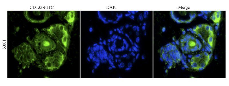
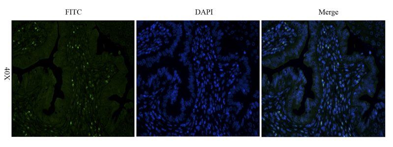
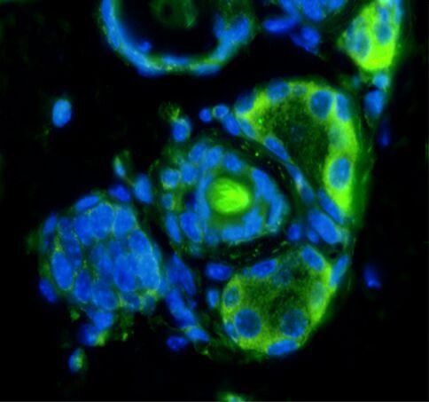
IF
Feitosa, Neli Patrícia Pereira et al. Cancerous and non-neoplastic stem cells in the stomach similarly express CD44 and CD133 Acta Histochem, 123, 151787 (2021)
Kase, Marju et al. Impact of CD133 positive stem cell proportion on survival in patients with glioblastoma multiforme Radiol Oncol, 47, 405-410 (2013)
Hirashima, Kanji et al. Cell biological profiling of reprogrammed cancer stem cell-like colon cancer cells maintained in culture Cell Tissue Res, 375, 697-707 (2019)
Nowacki, Maciej et al. Nanovehicles as a novel target strategy for hyperthermic intraperitoneal chemotherapy: a multidisciplinary study of peritoneal carcinomatosis Oncotarget, 6, 22776-22798 (2015)
Zou, Yuhong et al. Nrf2 is involved in maintaining hepatocyte identity during liver regeneration PLoS One, 9, e107423 (2014)
Coni, Pierpaolo et al. Exploring cell surface markers and cell-cell interactions of human breast milk stem cells J Public Health Res, 12, (2023)
Yang, Meng-Yin et al. An innovative three-dimensional gelatin foam culture system for improved study of glioblastoma stem cell behavior J Biomed Mater Res B Appl Biomater, 103, 618-628 (2015)
Zhu, Yifan et al. Influence of interferon-α on the expression of the cancer stem cell markers in pancreatic carcinoma cells Exp Cell Res, 324, 146-156 (2014)
Nadal, Rosa et al. CD133 expression in circulating tumor cells from breast cancer patients: potential role in resistance to chemotherapy Int J Cancer, 133, 2398-2407 (2013)
Tu, Zhenbo et al. CXCR4 is involved in CD133-induced EMT in non-small cell lung cancer Int J Oncol, 50, 505-514 (2017)
Applications
IHC
Reactivity
Human
Sun, Zhonghai et al. Tumour stem cell markers CD133 and CD44 are useful prognostic factors after surgical resection of pancreatic neuroendocrine tumours Oncol Lett, 20, 341 (2020)
Applications
IHC
Reactivity
Human
Jensen, Todd et al. Descriptive analysis of tumor cells with stem like phenotypes in metastatic and benign adrenal tumors J Pediatr Surg, 50, 1493-1501 (2015)

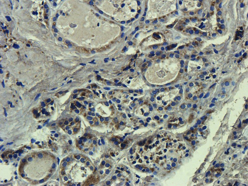
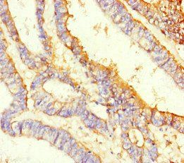
Immunohistochemical analysis of paraffin-embedded human mammary cancer tissue using CD133 antibody

IHC-P analysis of mouse heart tissue using CD133 antibody (Primary antibody diluted to 1:100)

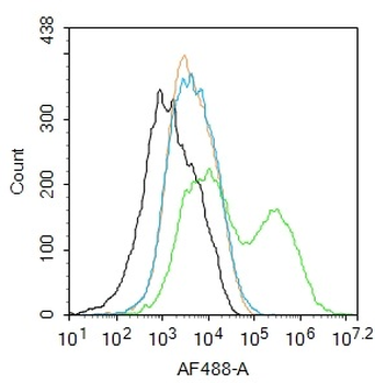
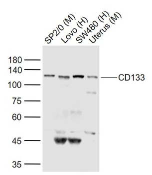
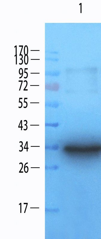
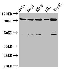
Western blot analysis of human cancer cell lysates using CD133 antibody [Reviews]

WB analysis of rat small intestine tissue using CD133 antibody (dilution at 1:200-1:1000 based on 1mg/mL)

Western Blot on human cells using CD133 antibody (Protocol steps under Review A) [Reviews]

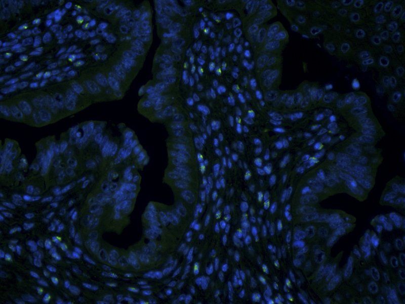
IF analysis analysis of mouse lung tissue using CD133 antibody (Dilution of primary antibody 1:200)

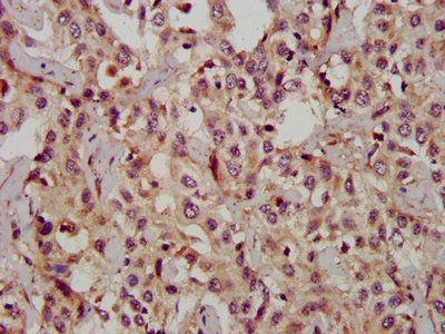
IHC-P analysis of mouse liver tissue using CD133 antibody (Dilution of primary antibody 1:100)

IHC-P analysis of mouse liver tissue using CD133 antibody (Primary antibody at 1:100)

Immunohistochemical staining of paraffin embedded human breast cancer tissue using anti-CD206 (primary antibody at 1:200)

IHC-P analysis of mouse heart tissue using CD133 antibody (Primary antibody at 1:100)

IHC-P analysis of mouse brain tissue using CD133 antibody (Dilution of primary antibody 1:100)

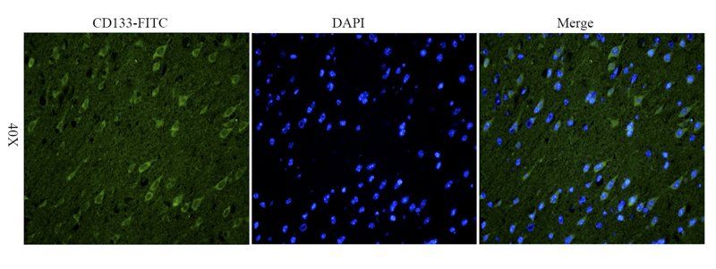
Immunofluorescent analysis of mouse brain tissue using CD133 antibody (Primary antibody diluted to 1:200)

Immunohistochemical analysis of formalin-fixed and paraffin embedded mouse skin tissue using CD133 antibody (Dilution of primary antibody 1:100)

IHC-P analysis of human lung cancer tissue using CD133 antibody (Dilution of primary antibody 1:100)

Immunohistochemical analysis of formalin-fixed and paraffin embedded human muscle tissue using CD133 antibody (Primary antibody diluted to 1:200)

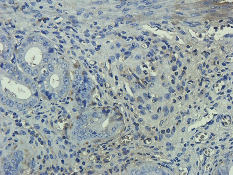
IHC-P analysis of human colonic adenocarcinoma tissue using CD133 antibody (Primary antibody at 1:100)

IHC-P analysis of human muscle tissue using CD133 antibody (Primary antibody at 1:100)

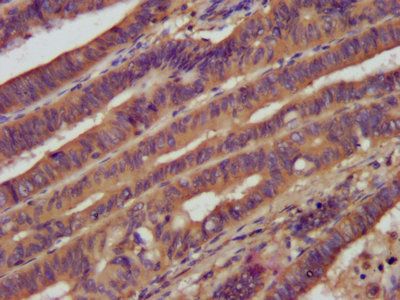
Immunohistochemical analysis of formalin-fixed and paraffin embedded human colonic adenocarcinoma tissue using CD133 antibody (Dilution of primary antibody 1:100)

IHC-P analysis of human lung cancer tissue using CD133 antibody (Primary antibody diluted to 1:100)

IHC-P analysis of human muscle tissue using CD133 antibody (Primary antibody diluted to 1:100)

IHC-P analysis of human colonic adenocarcinoma tissue using CD133 antibody (Primary antibody at 1:100)

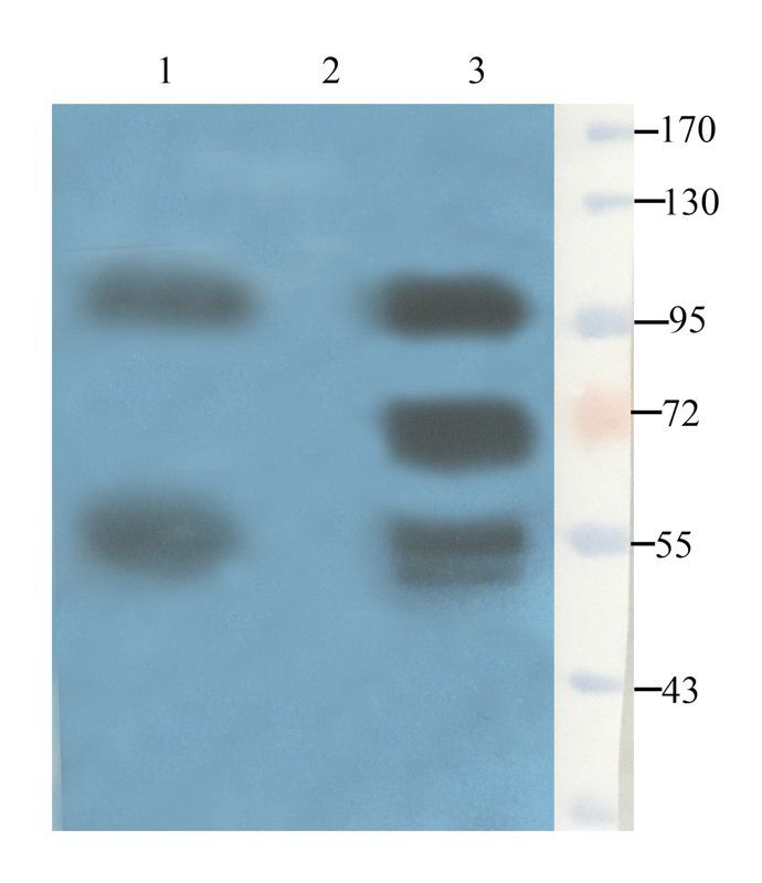
Western blot analysis of human muscle (Lane1), Rat muscle (Lane2), Rat bladder (Lane3) using CD133 antibody (Dilution at: 1:500)

Western blot analysis of rat small intestine tissue using CD133 antibody (dilution at 1:200-1:1000 based on 1mg/mL)

Immunohistochemical staining of human mammary fibroma tissue using anti-CD206 (dilution of primary antibody - 1:100)

IHC-P image of human breast cancer tissue using anti-CD206 (dilution of primary antibody at 1:100)

Western blot analysis of Mouse large intestine (Lane 1), Mouse spinal cord (Lane 2) using CD133 antibody (Dilution of primary antibody 1:200)

Immunohistochemical staining of paraffin embedded human endometrial cancer tissue using anti-CD206 (primary antibody at 1:200)

IHC-P image of human endometrial cancer tissue using anti-CD206 (dilution of primary antibody at 1:100)

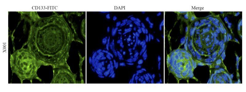
IF analysis of mouse skin tissue using CD133 antibody (Dilution of primary antibody 1:200)

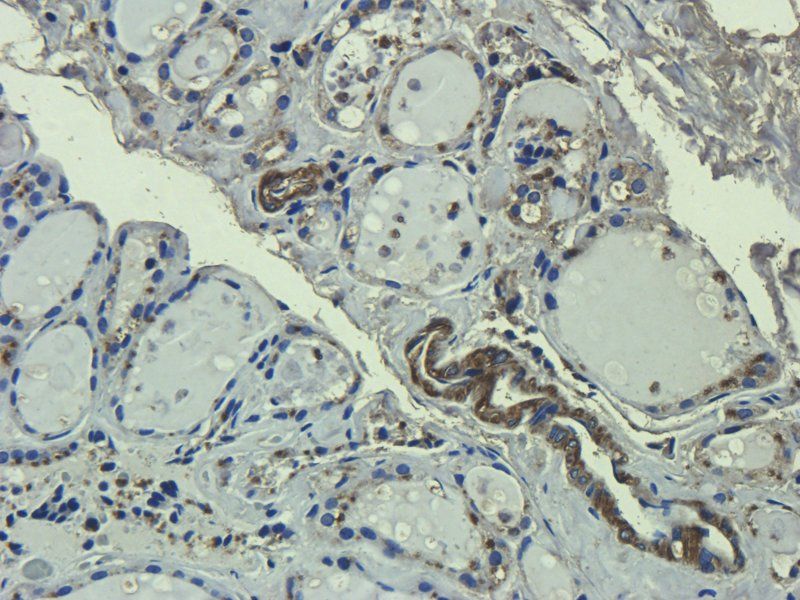
IHC-P analysis of mouse kidney tissue using CD133 antibody (Primary antibody diluted to 1:200)
- Item 1 of 9
- Item 1 of 5
CD133 Rabbit Polyclonal Antibody [orb526485]
FC, WB
Rat
Human, Mouse
Rabbit
Polyclonal
Unconjugated
200 μl, 100 μl, 50 μl - Item 1 of 5
- Item 1 of 6
CD133 / Prominin-1 (Intracellular domain) [orb1294396]
IF, IHC, WB
Human, Mouse, Rat
Rabbit
Polyclonal
Unconjugated
25 μl, 100 μl - Item 1 of 4


Submit a review
Filter by Rating

- 5 stars

- 4 stars
- 3 stars
- 2 stars
- 1 stars
CD133 antibody (orb99113)
We have used this CD133 antibody for quite some time, as it gives us highly reproducible results and easily identified staining. The concentration ratio allows us to do multiple sets of slides with the same lot and produces publication-quality results every time." Review date: 04 Sep 2020
- Catherine Langford
Rating
- 5 stars
