You have no items in your shopping cart.
Cart summary
Item 1 of 6
Item 1 of 6
CBS Antibody
Catalog Number: orb1263565

| Catalog Number | orb1263565 |
|---|---|
| Category | Antibodies |
| Description | CBS Antibody |
| Species/Host | Rabbit |
| Clonality | Polyclonal |
| Tested applications | FC, IF, IHC-P, WB |
| Predicted Reactivity | Monkey |
| Reactivity | Human, Rat |
| Isotype | Rabbit Ig |
| Immunogen | This CBS antibody is generated from rabbits immunized with a KLH conjugated synthetic peptide between 301-330 amino acids from the Central region of human CBS. |
| Antibody Type | Primary Antibody |
| Concentration | batch dependent |
| Form/Appearance | Liquid |
| Conjugation | Unconjugated |
| MW | 61 kDa |
| Target | CBS |
| UniProt ID | P35520 |
| NCBI | P35520 |
| Storage | Maintain refrigerated at 2-8°C for up to 2 weeks. For long term storage store at -20°C in small aliquots to prevent freeze-thaw cycles. |
| Buffer/Preservatives | Supplied in PBS with 0.09% (W/V) sodium azide. |
| Alternative names | Cystathionine beta-synthase, Beta-thionase, Serine Read more... |
| Note | For research use only |
| Application notes | For FACS starting dilution is: 1:25For WB starting dilution is: 1:2000For IHC-P starting dilution is: 1:25For IF starting dilution is: 1:10~50 |
| Expiration Date | 12 months from date of receipt. |

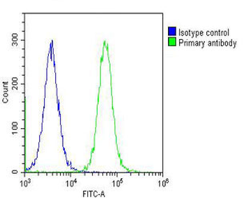
Overlay histogram showing Hela cells stained with Antibody (green line). The cells were fixed with 2% paraformaldehyde (10 min) and then permeabilized with 90% methanol for 10 min. The cells were then icubated in 2% bovine serum albumin to block non-specific protein-protein interactions followed by the antibody (1:25 dilution) for 60 min at 37°C. The secondary antibody used was Goat-Anti-Rabbit IgG, Conjugated Highly Cross-Adsorbed at 1/200 dilution for 40 min at 37°C. Isotype control antibody (blue line) was rabbit IgG (1ug/1x10^6 cells) used under the same conditions. Acquisition of > 10000 events was performed.

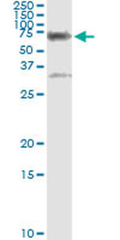
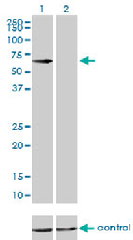
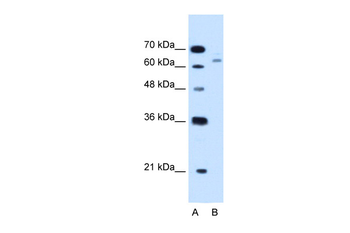
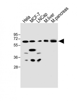
Western Blot at 1:2000 dilution + Hela whole cell lysate Lysates/proteins at 20 ug per lane.

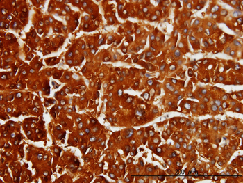
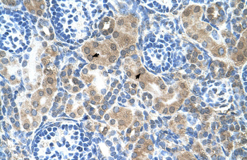
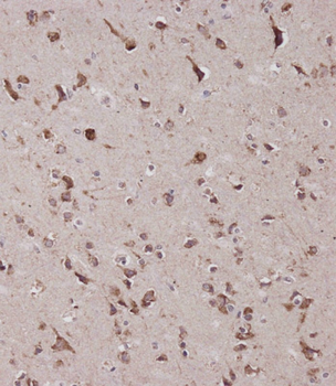
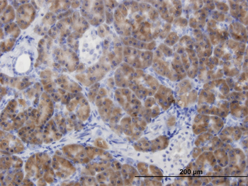
Immunohistochemical analysis of paraffin-embedded H. brain section using CBS Antibody. Antibody was diluted at 1:25 dilution. A peroxidase-conjugated goat anti-rabbit IgG at 1:400 dilution was used as the secondary antibody, followed by DAB staining.

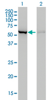
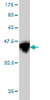
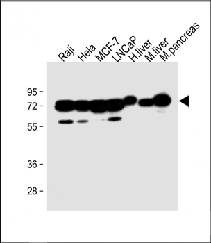
Western blot analysis in Raji cell line, rat brain and cerebellum tissue lysates (35 ug/lane).

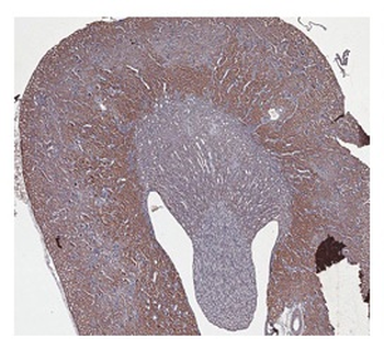
Formalin-fixed and paraffin-embedded human brain tissue reacted with CBS Antibody, which was peroxidase-conjugated to the secondary antibody, followed by DAB staining.

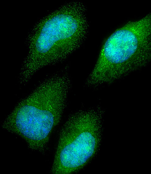
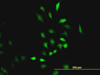
Confocal immunofluorescent analysis of CBS Antibody with 293 cell followed by Alexa Fluor 488-conjugated goat anti-rabbit lgG (green). Actin filaments have been labeled with Alexa Fluor 555 phalloidin (red).DAPI was used to stain the cell nuclear (blue).
- Item 1 of 8
CBS monoclonal antibody (M01), clone 3E1 [orb2294999]
ELISA, IHC-P, IP, WB
Human, Rat
Mouse
Monoclonal
Unconjugated
50 μg - Item 1 of 8
CBS Rabbit Polyclonal Antibody [orb579564]
IHC, WB
Bovine, Canine, Equine, Guinea pig, Rabbit, Yeast, Zebrafish
Human, Mouse, Rat
Rabbit
Polyclonal
Unconjugated
100 μl - Item 1 of 5
CBS Antibody [orb401264]
ELISA, IF, IHC, IP, WB
Human, Mouse
Rabbit
Polyclonal
Unconjugated
50 μg, 100 μg - Item 1 of 6
CBS Antibody (Center) [orb1929685]
FC, IF, IHC-P, WB
Human, Mouse, Rat
Rabbit
Polyclonal
Unconjugated
100 μl, 50 μl - Item 1 of 4
CBS monoclonal antibody (M07), clone 5F7 [orb2294995]
ELISA, IF, IHC-P, WB
Human
Mouse
Monoclonal
Unconjugated
100 μg