You have no items in your shopping cart.
Cart summary
Item 1 of 6
Item 1 of 6
CBS Antibody (Center)
Catalog Number: orb1929685

| Catalog Number | orb1929685 |
|---|---|
| Category | Antibodies |
| Description | Affinity Purified Rabbit Polyclonal Antibody (Pab) |
| Species/Host | Rabbit |
| Clonality | Polyclonal |
| Clone Number | RB21183 |
| Tested applications | FC, IF, IHC-P, WB |
| Reactivity | Human, Mouse, Rat |
| Isotype | Rabbit IgG |
| Dilution range | WB: 1:500, IF: 1:25, WB: 1:2000, IHC-P-Leica: 1:500, IHC-P-Leica: 1:500, FC: 1:25 |
| Form/Appearance | Purified polyclonal antibody supplied in PBS with 0.09% (W/V) sodium azide. This antibody is purified through a protein A column, followed by peptide affinity purification. |
| Conjugation | Unconjugated |
| MW | 60587 Da |
| Target | This CBS antibody is generated from rabbits immunized with a KLH conjugated synthetic peptide between 301-330 amino acids from the Central region of human CBS. |
| UniProt ID | P35520 |
| NCBI | NP_000062.1, NP_001171479.1, NP_001171480.1 |
| Storage | Maintain refrigerated at 2-8°C for up to 2 weeks. For long term storage store at -20°C in small aliquots to prevent freeze-thaw cycles |
| Alternative names | Cystathionine beta-synthase, Beta-thionase, Serine Read more... |
| Note | For research use only |
| Expiration Date | 12 months from date of receipt. |

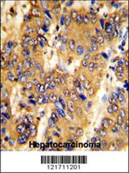
Immunohistochemical analysis on paraffin-embedded Human liver tissue was performed on the Leica BOND RXm. Tissue was fixed with formaldehyde at room temperature. Heat induced epitope retrieval was performed by EDTA buffer (pH9.0). Samples were incubated with primary antibody (1:500) for 15 min at room temperature. Leica Bond Polymer Refine Detection was used as the secondary antibody.
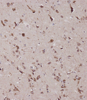
Immunohistochemical analysis on paraffin-embedded Human brain tissue was performed on the Leica BOND RXm. Tissue was fixed with formaldehyde at room temperature. Heat induced epitope retrieval was performed by EDTA buffer (pH9.0). Samples were incubated with primary antibody (1:500) for 15 min at room temperature. Leica Bond Polymer Refine Detection was used as the secondary antibody.
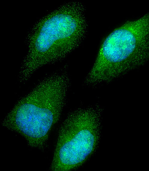
Immunofluorescent analysis of 4% paraformaldehyde-fixed, 0.1% Triton X-100 permeabilized HeLa (human cervical epithelial adenocarcinoma cell line) cells labeling CBS at 1/25 dilution, followed by Dylight 488-conjugated goat anti-rabbit IgG secondary antibody at 1/200 dilution (green). Immunofluorescence image showing cytoplasm and nucleus staining on HeLa cell line. The nuclear counter stain is DAPI (blue).
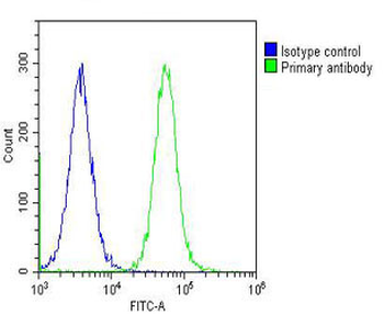
Overlay histogram showing Hela cells stained (green line). The cells were fixed with 2% paraformaldehyde (10 min) and then permeabilized with 90% methanol for 10 min. The cells were then icubated in 2% bovine serum albumin to block non-specific protein-protein interactions followed by the antibody (1:25 dilution) for 60 min at 37°C. The secondary antibody used was Goat-Anti-Rabbit IgG, DyLight 488 Conjugated Highly Cross-Adsorbed at 1/200 dilution for 40 min at 37°C. Isotype control antibody (blue line) was rabbit IgG (1 μg/1x10^6 cells) used under the same conditions. Acquisition of > 10000 events was performed.
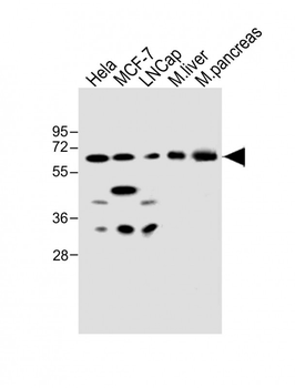
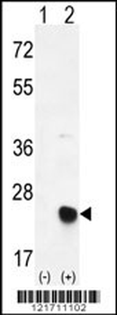
All lanes: Anti-CBS Antibody (Center) at 1:500 dilution. Lane 1: Hela whole cell lysate. Lane 2: MCF-7 whole cell lysate. Lane 3: LNCap whole cell lysate. Lane 4: mouse liver lysate. Lane 5: mouse pancreas lysate. Lysates/proteins at 20 µg per lane. Secondary Goat Anti-Rabbit IgG, (H+L), Peroxidase conjugated at 1/10000 dilution. Predicted band size: 61 kDa. Blocking/Dilution buffer: 5% NFDM/TBST.
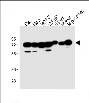
All lanes: Anti-CBS Antibody (Center) at 1:2000 dilution. Lane 1: Raji whole cell lysate. Lane 2: Hela whole cell lysate. Lane 3: MCF-7 whole cell lysate. Lane 4: LNCaP whole cell lysate. Lane 5: Human liver lysate. Lane 6: Mouse liver lysate. Lane 7: Mouse pancreas lysate. Lysates/proteins at 20 µg per lane. Secondary Goat Anti-Rabbit IgG, (H+L), Peroxidase conjugated at 1/10000 dilution. Predicted band size: 61 kDa. Blocking/Dilution buffer: 5% NFDM/TBST.
- Item 1 of 3

CBS Antibody (Center) [orb1166443]
FC, IF, IHC-P, WB
Human, Rat
Rabbit
Polyclonal
Unconjugated
100 μl, 30 μl