You have no items in your shopping cart.
Cart summary
Item 1 of 3
Item 1 of 3
Cbl-c antibody
Catalog Number: orb345519

| Catalog Number | orb345519 |
|---|---|
| Category | Antibodies |
| Description | Cbl-c antibody |
| Species/Host | Rabbit |
| Clonality | Polyclonal |
| Tested applications | ELISA, IHC, IP, WB |
| Reactivity | Human |
| Isotype | IgG |
| Immunogen | This affinity-purified antibody was prepared from whole rabbit serum produced by repeated immunizations with a synthetic peptide corresponding to the C-Terminal portion of Human Cbl-c. |
| Concentration | 1.2 mg/ml |
| Dilution range | ELISA: 1:10,000 - 1:50,000, IHC: 1:500 - 1:3,000, IP: User Optimized, WB: 1:500 - 1:3,000 |
| Form/Appearance | Liquid (sterile filtered) |
| Purity | This affinity purified antibody is directed against human Cbl-c protein. The product was affinity purified from monospecific antiserum by immunoaffinity purification. A BLAST analysis was used to suggest that this antibody would react with Cbl-c from human and chimpanzee sources. Expect partial reactivity against mouse and rat sources of Cbl-c as ~83% sequence homology is on record for the immunogen sequence. Reactivity with Cbl-c from other sources has not been determined. No reactivity is expected with Cbl-a or Cbl-b. |
| Conjugation | Unconjugated |
| UniProt ID | Q9ULV8 |
| NCBI | 125987803 |
| Storage | Store vial at -20° C prior to opening. Aliquot contents and freeze at -20° C or below for extended storage. Avoid cycles of freezing and thawing. Centrifuge product if not completely clear after standing at room temperature. This product is stable for several weeks at 4° C as an undiluted liquid. Dilute only prior to immediate use. |
| Buffer/Preservatives | 0.01% (w/v) Sodium Azide |
| Alternative names | rabbit anti-Cbl-c Antibody, E3 ubiquitin-protein l Read more... |
| Note | For research use only |
| Application notes | This affinity purified antibody has been tested for use in ELISA, immunohistochemistry, immunoprecipitation and western blot. Specific conditions for reactivity should be optimized by the end user. Expect a band at ~52 kDa in size corresponding to Cbl-c by western blotting in the appropriate cell lysate or extract. |
| Expiration Date | 12 months from date of receipt. |
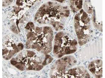
Biorbyt's Affinity Purified anti-Cbl-c antibody shows strong nuclear and cytoplasmic staining of cells in tubuli in human kidney tissue. Tissue was formalin-fixed and paraffin embedded. Brown color indicates presence of protein, blue color shows cell nuclei.
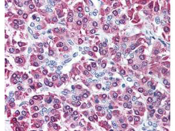
Biorbyt's affinity purified anti-Cbl-c antibody was used at 5 µg/ml to detect signal in a variety of tissues including multi-human, multi-brain and multi-cancer slides. This image shows moderate intracellular positive staining of human pancreatic acinar epithelium at 40X. Tissue was formalin-fixed and paraffin embedded. The image shows localization of the antibody as the precipitated red signal, with a hematoxylin purple nuclear counterstain.
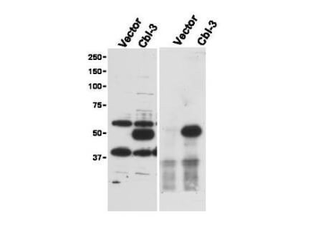
Immunoprecipitation (right) and western blot (left) using Biorbyt's Affinity Purified anti-Cbl-c antibody shows detection of a predominant band at ~52 kDa corresponding to Cbl-c. Lysates used are from HEK293T cells transfected with empty vector or with Cbl-c and western blotting (left panel). The predicted size of Cbl-c is 52 kDa. Size markers in kDa are shown to the left of the panel. The (right panel) shows immunoprecipitation with Rabbit anti-Cbl-c followed by western blotting using a Goat anti-Cbl-c antibody.
- Item 1 of 4
CBL antibody [orb10856]
ICC, IF, IHC, IP, WB
Human, Mouse, Rat
Polyclonal
Unconjugated
50 μl, 100 μl, 200 μl - Item 1 of 3
- Item 1 of 2
- Item 1 of 2
- Item 1 of 2


Submit a review
Filter by Rating

- 5 stars

- 4 stars
- 3 stars
- 2 stars
- 1 stars
