You have no items in your shopping cart.
Cart summary
Item 1 of 7
Item 1 of 7
CASZ1 antibody
Catalog Number: orb345676

| Catalog Number | orb345676 |
|---|---|
| Category | Antibodies |
| Description | CASZ1 antibody |
| Species/Host | Rabbit |
| Clonality | Polyclonal |
| Tested applications | ELISA, IF, IHC, WB |
| Reactivity | Human |
| Isotype | IgG |
| Immunogen | This affinity purified antibody was prepared from whole rabbit serum produced by repeated immunizations with a synthetic peptide corresponding to an internal region of Human Casz1 protein. |
| Concentration | 1.0 mg/mL |
| Dilution range | ELISA: 1:45,000, IHC: User Optimized, IF: User Optimized, WB: 1:1,000 - 1:5,000 |
| Form/Appearance | Liquid (sterile filtered) |
| Purity | This affinity purified antibody is directed against human Casz1 protein. The product was affinity purified from monospecific antiserum by immunoaffinity purification. A BLAST analysis was used to suggest reactivity with CASZ1 proteins from human, mouse, Drosophila, chimpanzee, and macaque based on a 100% homology. Partial reactivity is expected with horse and dog CASZ1 based on a 92% homology with the immunizing sequence. Cross-reactivity with CASZ1 from other sources has not been determined. |
| Conjugation | Unconjugated |
| UniProt ID | Q86V15 |
| NCBI | 145207289 |
| Storage | Store vial at -20° C or below prior to opening. This vial contains a relatively low volume of reagent (25 µL). To minimize loss of volume dilute 1:10 by adding 225 µL of the buffer stated above directly to the vial. Recap, mix thoroughly and briefly centrifuge to collect the volume at the bottom of the vial. Use this intermediate dilution when calculating final dilutions as recommended below. Store the vial at -20°C or below after dilution. Avoid cycles of freezing and thawing. |
| Buffer/Preservatives | 0.01% (w/v) Sodium Azide |
| Alternative names | rabbit anti-CASZ1 Antibody, CASZ1, Zinc finger pro Read more... |
| Note | For research use only |
| Application notes | This antibody has been tested for use in ELISA, IHC, IF, and western blotting. This antiserum detects endogenous Casz1 proteins, both the hCasz5 and hCasz11 isoforms. Expect a band approximately 125 kDa for hCasz5 and 190 kDa for hCaz11 in size corresponding Casz1 by western blotting in the appropriate cell lysate or extract. Specific conditions for reactivity should be optimized by the end user. |
| Expiration Date | 12 months from date of receipt. |
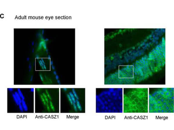
Immunofluorescence of Rabbit anti-CASZ1 Antibody. Tissue: adult murine ocular tissue. Antibody: Rabbit Anti-CASZ1 Antibody. Counterstain: DAPI. Localization: nucleus in lens epithelia but primarily localizes in the cytoplasm in photoreceptor cells.
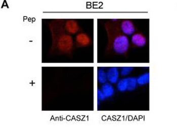
Immunofluorescence results of Endogenous CASZ1. Cells: BE2 cells. With or without Pre-Incubation of Anti-CASZ1 Antibody with CASZ1 Peptide. Staining: Rabbit Anti-CASZ1 Antibody. Chromatin counter stain: DAPI.

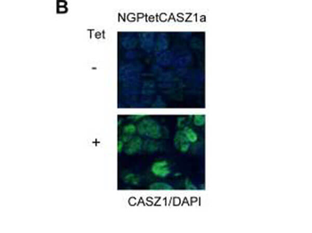
Immunofluorescence results of Rabbit Anti-CASZ1 Antibody. Tissue: Mouse Xenograft tumor of human NB cell line transfected with or without tetracycline inducible CASZ1 (NGPtetCASZ1a). Antibody: Rabbit Anti-CASZ1 Antibody. Counterstain: DAPI.
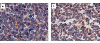
Immunohistochemistry results of Rabbit Anti-hCasz1 Antibody. Tissue: NB patient tumor. A. CASZ1 localized exclusively in the cytoplasm. B. CASZ1 localized in the cytoplasm and nucleus. Primary Antibody: Rabbit Anti-CASZ1 stained brown. Nucleus counterstained with hematoxylin (blue).

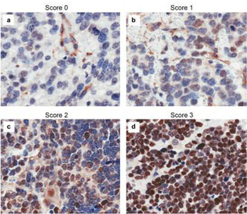
Immunohistochemistry results of Rabbit Anti-hCASZ1 Antibody. Tissue: NB patient tumor. A. Score 0- a rare positive nuclei. B. Score 1- (1-10% positive) equivocal/uninterpretable. C. Score 2- (10-50% positive) weak positive. D. Score 3- (> 50% positive) strong positive. Primary Antibody: Rabbit Anti-CASZ1 stained brown. Nucleus counterstained with hematoxylin (blue). Localization: Nuclear.
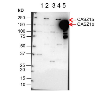
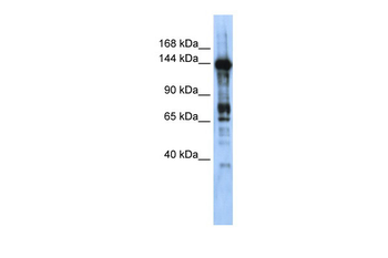
Western Blot of Anti-CASZ1 Antibody. Lane 1: NBLS Cytoplasmic (20 µg). Lane 2: NBLS Nuclear (3 µg). Lane 3: BE2C Cytoplasmic (30 µg). Lane 4: BE2C Nuclear (7 µg). Lane 5: SY5Y-CASZ1b (10 µg). Block: 5% Blotto/TTBS for 1 hour. Primary: Casz1 1:10000 for 1 hour. Secondary: Goat anti-Rabbit HRP for 1 hour. 240sec exposure. Detects nuclear endogenous CASZ1a and CASZ1b; and transiently transfected CASZ1b isoform.
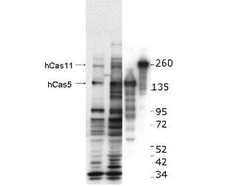
Western blot using Biorbyt's anti-hCASZ1 antibody. This blot shows detection of endogenous and transfected human CASZ1 protein in fresh whole cell lysate (~30 µg). Lane 1: BE2(s) cell lysate. Lane 2: BE2(N) cell lysate. Lane 3: SY5Y transfected with hCASZ5 (125 kDa). Lane 4: SY5Y transfected with hCASZ11 (190 kDa). Protein was resolved by SDS-PAGE and transferred onto nitrocellulose. After blocking, the membrane was probed with the primary antibody diluted to 1:1000 for 1.5 hours at room temperature then incubated with HRP-conjugated Goat Anti-Rabbit antibody for 45 min. at room temperature.
- Item 1 of 7
- Item 1 of 2
- Item 1 of 2
CASZ1 antibody [orb574530]
WB
Bovine, Equine, Guinea pig, Human, Rat
Mouse
Rabbit
Polyclonal
Unconjugated
100 μl - Item 1 of 1
- Item 1 of 1


Submit a review
Filter by Rating

- 5 stars

- 4 stars
- 3 stars
- 2 stars
- 1 stars
