You have no items in your shopping cart.
Cart summary
Item 1 of 4
Item 1 of 4
CaMKII Antibody: Biotin
Catalog Number: orb147058

| Catalog Number | orb147058 |
|---|---|
| Category | Antibodies |
| Description | Mouse monoclonal to CaMKII (Biotin). CaMKII is an important member of calcium/calmodulin- activated protein kinase family, functioning in neural synaptic stimulation and T-cell receptor signaling. CaMKII is expressed in many different tissues but is specifically found in the neurons of the forebrain and its mRNA is found within the dendrites and the soma of the neuron. The CaMKII that is found in the neurons consist of two subunits of 52 (termed alpha genes) and 60 kDa (beta genes). CaMKII has catalytic and regulatory domains, as well as an ATP-binding domain, and a consensus phosphorylation site (3-7). The binding of Ca2+ auto inhibitory effect and activates the kinase. /calmodulin to its regulatory domain releases its This kinase activation results in auto phosphorylation at threonine 286. The threonine phosphorylation state of CaMKII can be regulated through PP1/PKA. Whereas PP1 (protein phosphatase 1) dephosphorylates phospho-CaMKII at Thr286, PKA (protein kinase A) prevents this dephosphorylation. Auto phosphorylation also enables CaMKII to attain an enhanced affinity for NMDA receptors in postsynaptic densities (10-12).. |
| Species/Host | Mouse |
| Clonality | Monoclonal |
| Clone Number | 22B1 |
| Tested applications | ELISA, ICC, IF, IHC, WB |
| Reactivity | Human, Mouse, Rat |
| Isotype | IgG1 |
| Immunogen | Synthetic peptide |
| Concentration | 1 mg/ml |
| Dilution range | WB (1:1000), IHC (1:100), ICC/IF (1:1000) |
| Conjugation | Biotin |
| Target | CaMKII |
| Entrez | 25400 |
| UniProt ID | P11275 |
| NCBI | NP_037052.1 |
| Storage | Conjugated antibodies should be stored according to the product label |
| Buffer/Preservatives | 136.36mM Ethanolamine, and 9.55mM Sodium Bicarbonate in 95.45% PBS |
| Alternative names | CSAID Binding protein 1 antibody, CSBP1 antibody, Read more... |
| Note | For research use only |
| Application notes | 1 µg/ml was sufficient for detection of 0.2 µg CamKII by colorimetric immunoblot analysis using Goat Anti-Mouse IgG:HRP as the secondary. |
| Expiration Date | 12 months from date of receipt. |

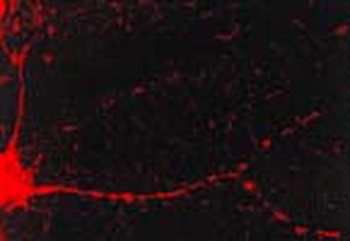
Immunocytochemistry/Immunofluorescence analysis using Mouse Anti-CaMKII Monoclonal Antibody, Clone 22B1. Tissue: dissociated hippocampal neurons. Species: Rat. Fixation: Cold 4% paraformaldehyde/0.2% glutaraldehyde in 0.1M sodium phosphate buffer. Primary Antibody: Mouse Anti-CaMKII Monoclonal Antibody at 1:1000 for 12 hours at 4°C. Secondary Antibody: FITC Goat Anti-Mouse IgG (green) at 1:50 for 30 minutes at RT. Magnification: 10X.
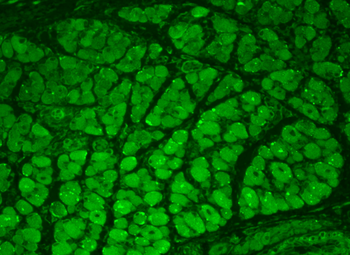
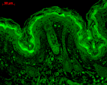
Immunohistochemistry analysis using Mouse Anti-CaMKII Monoclonal Antibody, Clone 22B1. Tissue: backskin. Species: Mouse. Fixation: Bouin's Fixative and paraffin-embedded. Primary Antibody: Mouse Anti-CaMKII Monoclonal Antibody at 1:100 for 1 hour at RT. Secondary Antibody: FITC Goat Anti-Mouse (green) at 1:50 for 1 hour at RT. Localization: Muscle, hair follicle, epidermis. Backskin obtained from transgenic mice.
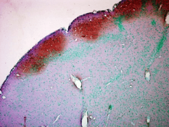
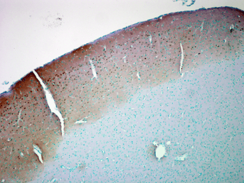
Immunohistochemistry analysis using Mouse Anti-CaMKII Monoclonal Antibody, Clone 22B1. Tissue: colon carcinoma. Species: Human. Fixation: Formalin. Primary Antibody: Mouse Anti-CaMKII Monoclonal Antibody at 1:5000 for 12 hours at 4°C. Secondary Antibody: Biotin Goat Anti-Mouse at 1:2000 for 1 hour at RT. Counterstain: Mayer Hematoxylin (purple/blue) nuclear stain at 200 μl for 2 minutes at RT. Magnification: 40x.
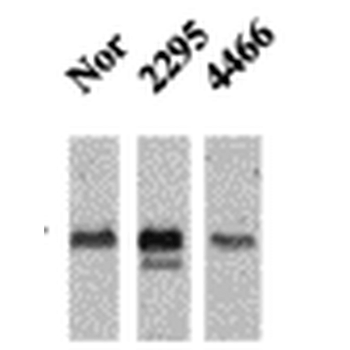
Western Blot analysis of Mouse Ventricle lysates showing detection of CaMKII protein using Mouse Anti-CaMKII Monoclonal Antibody, Clone 22B1. Primary Antibody: Mouse Anti-CaMKII Monoclonal Antibody at 1:1000. Analysis of CaMKII and NFAT phosphorylation in ventricles of 14 day old mice over-expressing CaMK.
- Item 1 of 5
CaMKII (Alpha-Specific) Antibody: Biotin [orb147041]
ELISA, ICC, IF, IHC, WB
Bovine, Human, Mouse, Rat
Mouse
Monoclonal
Biotin
100 μg - Item 1 of 1
Mouse Calcium/Calmodulin Dependent Protein Kinase II Gamma (CAMK2��) ELISA [orb1817294]
Mouse
0.16-10 ng/mL
0.081 ng/mL
96 T, 48 T 
CaMKII delta Rabbit Polyclonal Antibody (Biotin) [orb445431]
WB
Bovine, Porcine, Rat
Mouse
Rabbit
Polyclonal
Biotin
100 μlCaMK2 alpha Rabbit Polyclonal Antibody (Biotin) [orb457248]
ICC, IF, IHC-Fr, IHC-P, WB
Human, Mouse, Rat
Rabbit
Polyclonal
Biotin
100 μl