You have no items in your shopping cart.
Calreticulin Antibody: Biotin
Catalog Number: orb151513
| Catalog Number | orb151513 |
|---|---|
| Category | Antibodies |
| Description | Rabbit polyclonal to Calreticulin (Biotin). Calreticulin is a multifunctional, highly conserved Ca2+ -binding protein that is localized to the endoplasmic reticulum (ER), but has also been detected in the nucleus and nuclear envelop. Like many other ER proteins, it has the conserved ER retention KDEL (Lys-Asp-Glu-Leu) sequence at its C-terminus (1-3). CRTs three domains include a 180 residue N-terminal domain, a proline-rich P-domain (residues 189-288) that binds Ca2+ with high affinity and shares homology with calnexin (CNX) and calmegin, and a 110 residue C-terminal domain that binds Ca2+ with low affinity but high capacity (1,3). Recent studies suggest that this soluble ER protein has a multifunctional role. It appears to be involved in calcium storage and regulation as well as having a molecular chaperone activity. It has been shown to interact with the cytoskeleton and to be involved in the regulation of gene expression. Calreticulin may also play a role in cellular proliferation including its apparent activity in the proliferation of certain viruses within mammalian host cells , and it has also been shown to be induced in response to various types of cell stress including amino acid deprivation. Close interconnections among protein synthesis, gene expression and calcium signaling have been observed by many researchers in recent years. Calreticulin might be centrally located and therefore it crucially participates in the coordination of many functions by the cell. Studies also suggest its involvement in a few diseases such as systemic lupus erythematosus, rheumatoid arthritis, celiac disease, complete congenital heart block, and halothane hepatitis.. |
| Species/Host | Rabbit |
| Clonality | Polyclonal |
| Tested applications | ELISA, ICC, IF, IHC, WB |
| Reactivity | Bovine, Canine, Gallus, Guinea pig, Hamster, Human, Monkey, Mouse, Porcine, Rabbit, Rat, Sheep |
| Immunogen | Human calreticulin synthetic peptide with a cysteine residue added and the peptide conjugated to KLH |
| Concentration | 1 mg/ml |
| Dilution range | WB (1:1000), IHC (1:100), ICC/IF (1:100) |
| Conjugation | Biotin |
| MW | 63kDa |
| Target | Calreticulin |
| Entrez | 811 |
| UniProt ID | P27797 |
| NCBI | NP_004334.1 |
| Storage | Conjugated antibodies should be stored according to the product label |
| Buffer/Preservatives | 136.36mM Ethanolamine, and 9.55mM Sodium Bicarbonate in 95.45% PBS |
| Alternative names | CALR antibody, Calregulin antibody, cC1qR antibody Read more... |
| Note | For research use only |
| Application notes | A 1:1000 dilution of SPC-122 was sufficient for detection of Calreticulin in 20 µg of HeLa cell lysate by ECL immunoblot analysis. |
| Expiration Date | 12 months from date of receipt. |

Immunocytochemistry/Immunofluorescence analysis using Rabbit Anti-Calreticulin Polyclonal Antibody. Tissue: Heat Shocked Cervical cancer cell line (HeLa). Species: Human. Fixation: 2% Formaldehyde for 20 min at RT. Primary Antibody: Rabbit Anti-Calreticulin Polyclonal Antibody at 1:100 for 12 hours at 4°C. Secondary Antibody: FITC Goat Anti-Rabbit (green) at 1:200 for 2 hours at RT. Counterstain: DAPI (blue) nuclear stain at 1:40000 for 2 hours at RT. Localization: Endoplasmic reticulum lumen. Cytoplasm. Magnification: 100x. (A) DAPI (blue) nuclear stain. (B) Anti-Calreticulin Antibody. (C) Composite. Heat Shocked at 42°C for 1h.

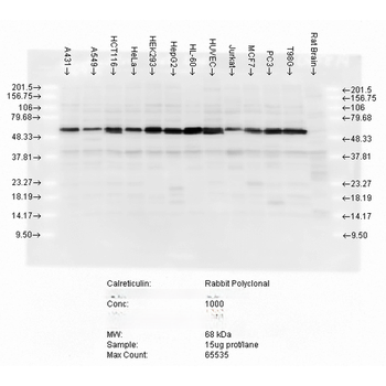
Western blot analysis of multiple cell lines lysates showing detection of Calreticulin protein using Rabbit Anti-Calreticulin Polyclonal Antibody. Load: 15 μgprotein. Block: 1.5% BSA for 30 minutes at RT. Primary Antibody: Rabbit Anti-Calreticulin Polyclonal Antibody at 1:5000 for 2 hours at RT. Secondary Antibody: Donkey Anti-Rabbit IgG: HRP for 1 hour at RT.

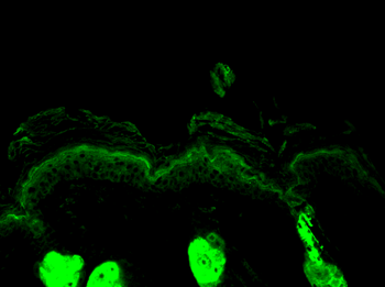
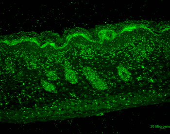
Immunohistochemistry analysis using Rabbit Anti-Calreticulin Polyclonal Antibody. Tissue: backskin. Species: Mouse. Fixation: Bouin's Fixative Solution. Primary Antibody: Rabbit Anti-Calreticulin Polyclonal Antibody at 1:100 for 1 hour at RT. Secondary Antibody: FITC Goat Anti-Rabbit (green) at 1:50 for 1 hour at RT. Localization: Cytoplasmic granule. Endoplasmic reticulum lumen.

Immunocytochemistry/Immunofluorescence analysis using Rabbit Anti-Calreticulin Polyclonal Antibody. Tissue: Heat Shocked Cervical cancer cell line (HeLa). Species: Human. Fixation: 2% Formaldehyde for 20 min at RT. Primary Antibody: Rabbit Anti-Calreticulin Polyclonal Antibody at 1:100 for 12 hours at 4°C. Secondary Antibody: R-PE Goat Anti-Rabbit (yellow) at 1:200 for 2 hours at RT. Counterstain: DAPI (blue) nuclear stain at 1:40000 for 2 hours at RT. Localization: Endoplasmic reticulum lumen. Cytoplasm. Magnification: 20x. (A) DAPI (blue) nuclear stain. (B) Anti-Calreticulin Antibody. (C) Composite. Heat Shocked at 42°C for 1h.
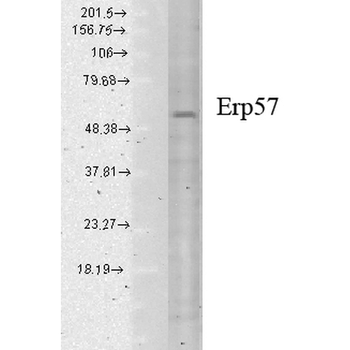
ERp57 (GRP58) Antibody: Biotin [orb147636]
ELISA, ICC, IF, IHC, WB
Bovine, Canine, Guinea pig, Hamster, Human, Monkey, Mouse, Porcine, Rabbit, Rat
Mouse
Monoclonal
Biotin
200 μg


