You have no items in your shopping cart.
Cart summary
Item 1 of 7
Item 1 of 7
CALR Antibody (Center)
Catalog Number: orb1929729

| Catalog Number | orb1929729 |
|---|---|
| Category | Antibodies |
| Description | Affinity Purified Rabbit Polyclonal Antibody (Pab) |
| Species/Host | Rabbit |
| Clonality | Polyclonal |
| Clone Number | RB21112 |
| Tested applications | FC, IHC-P, WB |
| Reactivity | Human, Rat |
| Isotype | Rabbit IgG |
| Dilution range | WB: 1:2000, WB: 1:1000, WB: 1:2000, WB: 1:1000, WB: 1:1000, IHC-P: 1:50~100, FC: 1:25 |
| Form/Appearance | Purified polyclonal antibody supplied in PBS with 0.09% (W/V) sodium azide. This antibody is purified through a protein A column, followed by peptide affinity purification. |
| Conjugation | Unconjugated |
| MW | 48142 Da |
| Target | This CALR antibody is generated from rabbits immunized with a KLH conjugated synthetic peptide between 277-305 amino acids from the Central region of human CALR. |
| UniProt ID | P27797 |
| NCBI | NP_004334.1 |
| Storage | Maintain refrigerated at 2-8°C for up to 2 weeks. For long term storage store at -20°C in small aliquots to prevent freeze-thaw cycles |
| Alternative names | Calreticulin, CRP55, Calregulin, Endoplasmic retic Read more... |
| Note | For research use only |
| Expiration Date | 12 months from date of receipt. |

Overlay histogram showing HeLa cells stained (green line). The cells were fixed with 2% paraformaldehyde (10 min) and then permeabilized with 90% methanol for 10 min. The cells were then icubated in 2% bovine serum albumin to block non-specific protein-protein interactions followed by the antibody (1:25 dilution) for 60 min at 37°C. The secondary antibody used was Goat-Anti-Rabbit IgG, DyLight 488 Conjugated Highly Cross-Adsorbed at 1/200 dilution for 40 min at 37°C. Isotype control antibody (blue line) was rabbit IgG1 (1 μg/1x10^6 cells) used under the same conditions. Acquisition of > 10000 events was performed.
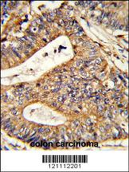
Formalin-fixed and paraffin-embedded human colon carcinoma reacted with CALR Antibody (Center), which was peroxidase-conjugated to the secondary antibody, followed by DAB staining. This data demonstrates the use of this antibody for immunohistochemistry; clinical relevance has not been evaluated.
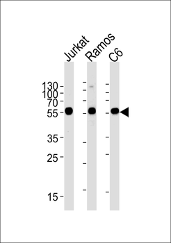
CALR Antibody (Center) western blot analysis in Jurkat, Ramos, rat C6 cell line lysates (35 ug/lane). This demonstrates the CALR antibody detected the CALR protein (arrow).
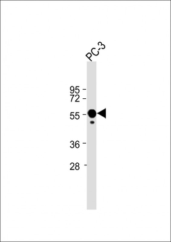
Anti-CALR Antibody (Center) at 1:2000 dilution + PC-3 whole cell lysate. Lysates/proteins at 20 µg per lane. Secondary Goat Anti-Rabbit IgG, (H+L), Peroxidase conjugated at 1/10000 dilution. Predicted band size: 55 kDa. Blocking/Dilution buffer: 5% NFDM/TBST.
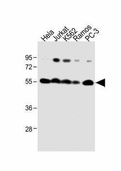
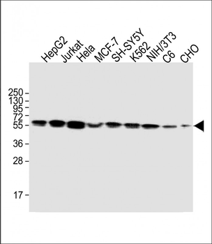
All lanes: Anti-CALR Antibody (Center) at 1:1000 dilution. Lane 1: Hela whole cell lysate. Lane 2: Jurkat whole cell lysate. Lane 3: K562 whole cell lysate. Lane 4: Ramos whole cell lysate. Lane 5: PC-3 whole cell lysate. Lysates/proteins at 20 µg per lane. Secondary Goat Anti-Rabbit IgG, (H+L), Peroxidase conjugated at 1/10000 dilution. Predicted band size: 48 kDa. Blocking/Dilution buffer: 5% NFDM/TBST.
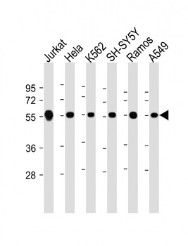
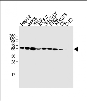
All lanes: Anti-CALR Antibody (Center) at 1:2000 dilution. Lane 1: Jurkat whole cell lysate. Lane 2: Hela whole cell lysate. Lane 3: K562 whole cell lysate. Lane 4: SH-SY5Y whole cell lysate. Lane 5: Ramos whole cell lysate. Lane 6: A549 whole cell lysate. Lysates/proteins at 20 µg per lane. Secondary Goat Anti-Rabbit IgG, (H+L), Peroxidase conjugated at 1/10000 dilution. Predicted band size: 48 kDa. Blocking/Dilution buffer: 5% NFDM/TBST.
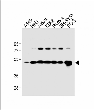
All lanes: Anti-CALR Antibody (Center) at 1:1000 dilution. Lane 1: A549 whole cell lysate. Lane 2: Hela whole cell lysate. Lane 3: Jurkat whole cell lysate. Lane 4: K562 whole cell lysate. Lane 5: Ramos whole cell lysate. Lane 6: SH-SY5Y whole cell lysate. Lane 7: PC-3 whole cell lysate. Lysates/proteins at 20 µg per lane. Secondary Goat Anti-Rabbit IgG, (H+L), Peroxidase conjugated at 1/10000 dilution. Predicted band size: 48 kDa. Blocking/Dilution buffer: 5% NFDM/TBST.
- Item 1 of 3