You have no items in your shopping cart.
Cart summary
Item 1 of 3
Item 1 of 3
c-FER antibody
Catalog Number: orb1473823

| Catalog Number | orb1473823 |
|---|---|
| Category | Antibodies |
| Description | Mouse monoclonal antibody to c-FER |
| Species/Host | Mouse |
| Clonality | Monoclonal |
| Tested applications | FC, IF, WB |
| Reactivity | Mouse |
| Immunogen | Recombinant fusion protein of human c-FER. The exact sequence is proprietary. |
| Dilution range | WB (1/500 - 1/2000), IF/IC (1/10 - 1/50), FC (1/10 - 1/50) |
| Form/Appearance | Mouse IgG2a kappa. Liquid in PBS, pH 7.3, 30% glycerol, and 0.01% sodium azide. |
| Conjugation | Unconjugated |
| Target | FER |
| Entrez | 14158 |
| UniProt ID | P70451 |
| Source | Mouse |
| Storage | Maintain refrigerated at 2-8°C for up to 2 weeks. For long term storage store at -20°C in small aliquots to prevent freeze-thaw cycles. |
| Buffer/Preservatives | Mouse IgG2a kappa. Liquid in PBS, pH 7.3, 30% glycerol, and 0.01% sodium azide. |
| Alternative names | TYK3; Tyrosine-protein kinase Fer; Feline encephal Read more... |
| Note | For research use only |
| Expiration Date | 12 months from date of receipt. |

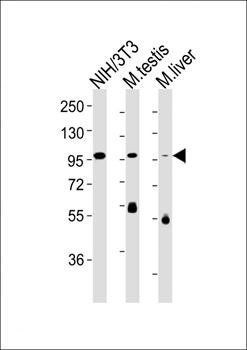
Western blot analysis of c-FER expression in NIH3T3 (A), mouse testis (B), mouse liver (C) whole cell lysates. (Predicted band size: 94 kD; Observed band size: 100 kD)

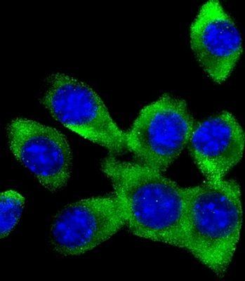
Immunofluorescent analysis of c-FER staining in NIH3T3 cells. Formalin-fixed cells were permeabilized with 0.1% Triton X-100 in TBS for 5-10 minutes and blocked with 3% BSA-PBS for 30 minutes at room temperature. Cells were probed with the primary antibody in 3% BSA-PBS and incubated overnight at 4 °C in a humidified chamber. Cells were washed with PBST and incubated with a AF488-conjugated secondary antibody (green) in PBS at room temperature in the dark. DAPI was used to stain the cell nuclei (blue).

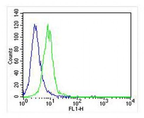
Flow cytometric analysis of NIH3T3 cells using Anti-c-FER Antibody. The cells were fixed with 2% paraformaldehyde (10 min) and then permeabilized with 90% methanol for 10 min. The cells were incubated in 2% bovine serum albumin to block non-specific protein-protein interactions followed by the antibody at 37 °C for 60 min. The secondary antibody Goat Anti-Mouse IgG (H&L) - AF488 was incubated at 37 °C for 40 min. Isotype control antibody (blue line) was used under the same condition.
- Item 1 of 4
- Item 1 of 4
- Item 1 of 3
- Item 1 of 3
- Item 1 of 2
FER (Phospho-Y402) antibody [orb393284]
IH, WB
Human, Mouse, Rat
Rabbit
Polyclonal
Unconjugated
30 μl, 100 μl, 200 μl


Submit a review
Filter by Rating

- 5 stars

- 4 stars
- 3 stars
- 2 stars
- 1 stars
