You have no items in your shopping cart.
Cart summary
Item 1 of 4
Item 1 of 4
BrdU antibody
Catalog Number: orb345680

| Catalog Number | orb345680 |
|---|---|
| Category | Antibodies |
| Description | BrdU antibody |
| Species/Host | Rabbit |
| Clonality | Polyclonal |
| Tested applications | ELISA, FC, IF, IHC, IP, WB |
| Reactivity | Other |
| Isotype | IgG |
| Immunogen | Anti-BrdU affinity purified antibody was purified from monospecific rabbit antiserum prepared via repeated immunizations with BromodeoxyUridine-KLH. |
| Concentration | 1.1 mg/mL |
| Dilution range | ELISA: 1:2000 - 1:10,000, FC: 1:50-1:100, IHC: 1:100-1:500, IF: 1:500, IP: User Optimized, WB: User Optimized |
| Form/Appearance | Liquid (sterile filtered) |
| Purity | Anti-BrdU Antibody was affinity purified from monospecific antiserum by immunoaffinity chromatography. |
| Conjugation | Unconjugated |
| Storage | Store BrdU Antibody at -20° C prior to opening. Aliquot contents and freeze at -20° C or below for extended storage. Avoid cycles of freezing and thawing. Centrifuge product if not completely clear after standing at room temperature. This product is stable for several weeks at 4° C as an undiluted liquid. Dilute only prior to immediate use. |
| Buffer/Preservatives | 0.01% (w/v) Sodium Azide |
| Alternative names | rabbit anti-BrdU Antibody, bromodeoxyuridine, 5-br Read more... |
| Note | For research use only |
| Application notes | Anti-BrdU Antibody has been tested by ELISA, WB, IHC, and is suitable for immunofluorescence microscopy and flow cytometry. Specific conditions for reactivity should be optimized by the end user. Expect to detect incorporated BrdU thymidine analog from replicated cells. |
| Expiration Date | 12 months from date of receipt. |

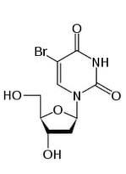
Bromodeoxyuridine (BrdU) chemical structure representation.
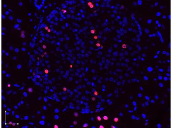
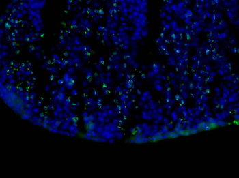
Immunofluorescence microscopy images of paraformaldehyde-fixed, paraffin-embedded pancreas sections stained with antibodies against BrdU (red or pink) and counterstained with DAPI (blue) and imaged with a 40× objective. DAPI stained nuclei (blue) indicate non-dividing cells, immunostained red and pink nuclei indicate actively dividing pancreatic ß-cells. The antibodies were diluted to 2.7 µg/ml. and incubated with tissue sections overnight at 4 degrees. Donkey anti-rabbit secondary antibody was diluted 1:2500.

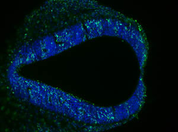
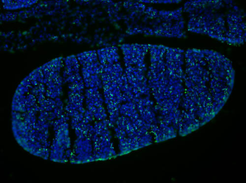
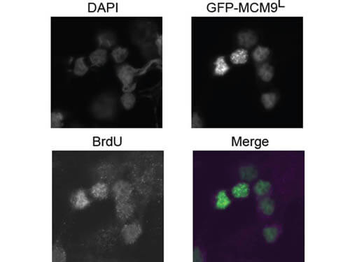
Immunofluorescence Microscopy of Rabbit anti-BrdU antibody. Tissue: 293T cells transfected expressing GFP-MCM9 L. Fixation: 0.5% PFA. Antigen retrieval: not required. Primary antibody: BrdU antibody at 10 µg/mL for 1 h at RT. Secondary antibody: Anti-rabbit ATTO550 secondary antibody at 1:10000 for 45 min at RT. Localization: BrdU is nuclear. Staining: BrdU in merged image shows with green and purple fluorescent signal with DAPI nuclear counterstain.
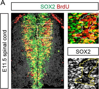
SOX2 represses proliferation in the developing spinal cord and stomach. (A) Percentage of cells expressing high or low levels of SOX2 labelled by a one hour pulse of BrdU in the E11.5 mouse spinal cord. Dotted lines in insets surround area of greatest BrdU incorporation. (B) Average background normalized SOX2 expression level and percentage of cells labelled by a one hour pulse of BrdU in the E11.5, E13.5 and E15.5 anterior and posterior stomach. (C) Percentage of electroporated cells in the chick spinal cord labelled by a 30 minute pulse of BrdU following misexpression of GFP, SOX2 or dnSOXB1. (D) Percentage of electroporated cells in E13.5 stomach explants labelled by a 30 minute pulse of BrdU following overexpression of GFP, SOX2 or dnSOXB1. All error bars represent standard deviations between experiments and p-values are calculated with two sided, unpaired t-tests (* = P< 0.05, ** = P< 0.01, *** = p< 0.001).
- Item 1 of 6
- Item 1 of 5
- Item 1 of 4
- Item 1 of 3
- Item 1 of 3


![BrdU Antibody [BRD469]](/images//pub/media/catalog/product/NewWebsite/15/orb1252671_1.jpg)
![BrdU Antibody [BRD469]](/images/pub/media/catalog/product/NewWebsite/15/orb1252671_2.jpg)
![BrdU Antibody [BRD469]](/images/pub/media/catalog/product/NewWebsite/15/orb1252671_3.jpg)
Submit a review
Filter by Rating

- 5 stars

- 4 stars
- 3 stars
- 2 stars
- 1 stars
