You have no items in your shopping cart.
Cart summary
Item 1 of 5
Item 1 of 5
BrdU antibody
Catalog Number: orb344495

| Catalog Number | orb344495 |
|---|---|
| Category | Antibodies |
| Description | BrdU antibody |
| Species/Host | Mouse |
| Clonality | Monoclonal |
| Clone Number | 29G6.E8 |
| Tested applications | DOT, ELISA, FC, IF, IHC, WB |
| Reactivity | Other |
| Isotype | IgG |
| Immunogen | Anti-BrdU monoclonal antibody was produced in mice by repeated immunizations prepared via immunizations with BromodeoxyUridine-KLH followed by hybridoma development. |
| Concentration | 1.0 mg/ml |
| Dilution range | ELISA: 1:2000 - 1:10000, FC: 1:50-1:100, IHC: 1:100-1:500, IF: 1:500-1:3000, WB: 1:2000 - 1:5000 |
| Form/Appearance | Liquid (sterile filtered) |
| Purity | Anti-BrdU Monoclonal Antibody was purified from ascites fluid by Protein A chromatography. This antibody reacts strongly with BrdU. Cross-reactivity is observed with CldU and IdU. |
| Conjugation | Unconjugated |
| Storage | Store BrdU Antibody at -20° C prior to opening. Aliquot contents and freeze at -20° C or below for extended storage. Avoid cycles of freezing and thawing. Centrifuge product if not completely clear after standing at room temperature. This product is stable for several weeks at 4° C as an undiluted liquid. Dilute only prior to immediate use. |
| Buffer/Preservatives | 0.01% (w/v) Sodium Azide |
| Alternative names | mouse anti-BrdU Antibody, Bromodeoxyuridine, 5-bro Read more... |
| Note | For research use only |
| Application notes | Anti-BrdU Antibody has been tested as suitable for immunofluorescence and immunoblot assays. Antibody may be suitable for additional immunoassays including flow cytometry and immunohistochemistry. Specific conditions for reactivity should be optimized by the end user. Antibody will detect incorporated BrdU thymidine analog from replicated cells. |
| Expiration Date | 12 months from date of receipt. |

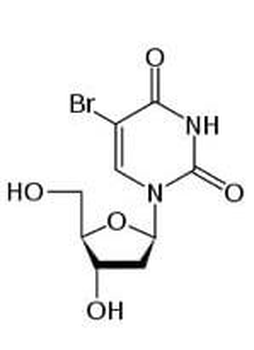
Bromodeoxyuridine (BrdU) chemical structure representation.
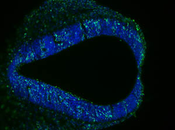
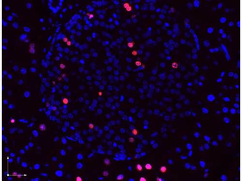
Immunofluorescence Microscopy of Mouse Anti-BrdU antibody. Tissue: OCT-embedded E10.5 mouse embryo. Localization: 20X, section through the developing hindbrain. Fixation: 4% PFA. Antigen retrieval: not required. Primary antibody: BrdU antibody at 1:1000 for overnight at 4°C in 0.4% PBS+ Triton with 1% normal sheep serum. Secondary antibody: Alexa Fluor 488 Anti-Mouse secondary antibody at 1:200 for 45 min at RT. Staining: Double labeled (green/blue) cells represent cells that were actively dividing.
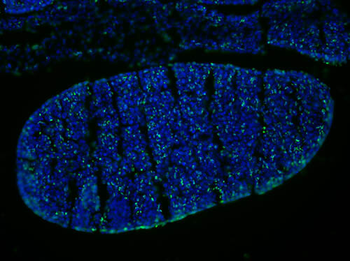
Immunofluorescence Microscopy of Mouse Anti-BrdU antibody. Tissue: OCT-embedded E10.5 mouse embryo. Localization: 20X, section through the developing limb bud. Fixation: 4% PFA. Antigen retrieval: not required. Primary antibody: BrdU antibody at 1:1000 in 0.4% PBS+ Triton with 1% normal sheep serum overnight at 4°C. Secondary antibody: Alexa Fluor 488 Anti-Mouse secondary antibody at 1:200 for 45 min at RT. Staining: Double labeled (green/blue) cells represent cells that were actively dividing.
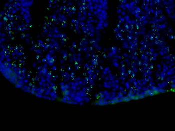
Immunofluorescence Microscopy of Mouse Anti-BrdU antibody. Tissue: OCT-embedded E10.5 mouse embryo. Localization: 40X, section through the developing limb bud. Fixation: 4% PFA. Antigen retrieval: not required. Primary antibody: BrdU antibody at 1:500 in 0.4% PBS+ Triton with 1% normal sheep serum overnight at 4°C. Secondary antibody: Alexa Fluor 488 Anti-Mouse secondary antibody at 1:200 for 45 min at RT. Staining: Double labeled (green/blue) cells represent cells that were actively dividing.
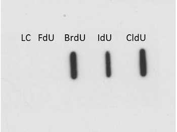
Western blot of Anti-BrdU antibody. Lane 1: loading control. Lane 2: FdU. Lane 3: BrdU. Lane 4: IdU. Lane 5 CldU. Load: 20 µg per lane. Primary antibody: Anti-BrdU antibody at 1:1000 for overnight at 4°C. Secondary antibody: IRDye800™ mouse secondary antibody at 1:10000 for 45 min at RT. Block: 5% BLOTTO overnight at 4°C. Predicted: BrdU. Other band(s): cross reactive bands observed for other nucleoside analogs, IdU and CldU.
- Item 1 of 6
- Item 1 of 4
- Item 1 of 4
- Item 1 of 3
- Item 1 of 3


![BrdU Antibody [BRD469]](/images//pub/media/catalog/product/NewWebsite/15/orb1252671_1.jpg)
![BrdU Antibody [BRD469]](/images/pub/media/catalog/product/NewWebsite/15/orb1252671_2.jpg)
![BrdU Antibody [BRD469]](/images/pub/media/catalog/product/NewWebsite/15/orb1252671_3.jpg)
Submit a review
Filter by Rating

- 5 stars

- 4 stars
- 3 stars
- 2 stars
- 1 stars
