You have no items in your shopping cart.
Cart summary
Item 1 of 4
Item 1 of 4
BNIP3 Antibody
Catalog Number: orb1269103

| Catalog Number | orb1269103 |
|---|---|
| Category | Antibodies |
| Description | BNIP3 Antibody |
| Species/Host | Rabbit |
| Clonality | Polyclonal |
| Tested applications | IF, IHC-P, WB |
| Reactivity | Human, Mouse |
| Isotype | Rabbit Ig |
| Immunogen | This BNIP3 antibody is generated from rabbits immunized with a KLH conjugated synthetic peptide between 152-187 amino acids from human BNIP3. |
| Antibody Type | Primary Antibody |
| Concentration | batch dependent |
| Form/Appearance | Liquid |
| Conjugation | Unconjugated |
| MW | 22 kDa |
| Target | BNIP3 |
| UniProt ID | Q12983 |
| NCBI | Q12983 |
| Storage | Maintain refrigerated at 2-8°C for up to 2 weeks. For long term storage store at -20°C in small aliquots to prevent freeze-thaw cycles. |
| Buffer/Preservatives | Supplied in PBS with 0.09% (W/V) sodium azide. |
| Alternative names | BCL2/adenovirus E1B 19 kDa protein-interacting pro Read more... |
| Note | For research use only |
| Application notes | For IF starting dilution is: 1:500For WB starting dilution is: 1:1000For IHC-P starting dilution is: 1:50~100 |
| Expiration Date | 12 months from date of receipt. |

Fluorescent confocal image of HepG2 cells stained with BNIP3 (BH3 Domain Specific) antibody. HepG2 cells were fixed with 4% PFA (20 min), permeabilized with Triton X-100 (0.2%, 30 min). Cells were then incubated with BNIP3 (BH3 Domain Specific) primary antibody (1:500, 2 h at room temperature). For secondary antibody, Alexa Fluor 488 conjugated donkey anti-rabbit antibody (green) was used (1:1000, 1h). Nuclei were counterstained with Hoechst 33342 (blue) (10 ug/ml, 5 min). BNIP3 immunoreactivity is localized to the cytoplasm of HepG2 cells.

Antibody is used in Western blot to detect NIP3 BH3 in Ramos cell lysate (lane 1) and in mouse brain tissue lysate (lane 2).

Freshly isolated mouse hepatocytes plated on coverslips (2 x10⁵ cells/22-mm glass coverslip) were cultured under normoxic conditions for 6 hr. The cells were then fixed in 2% paraformaldehyde in PBS for 1 hr, and processed for confocal immunofluorescence (red: F-actin, blue: ATP-synthase, green: BNIP3). Fluorescence labeling of BNIP3 accomplished with anti-BNIP3 antibody.

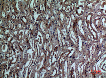
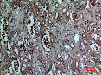
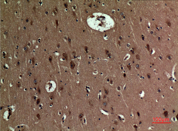
Formalin-fixed and paraffin-embedded human cancer tissue reacted with the primary antibody, which was peroxidase-conjugated to the secondary antibody, followed by DAB staining. BC = breast carcinoma; HC = hepatocarcinoma.
- Item 1 of 7
- Item 1 of 5
- Item 1 of 5
- Item 1 of 4
- Item 1 of 3