You have no items in your shopping cart.
Cart summary
Item 1 of 7
Item 1 of 7
BMI1 Antibody
Catalog Number: orb2643783

| Catalog Number | orb2643783 |
|---|---|
| Category | Antibodies |
| Description | The B cell-specific moloney murine leukemia virus integration site 1 (Bmi-1) is a transcriptional receptor of the polycomb gene family involved in several cellular processes including cell-cycle regulation, apoptosis, and maintenance of adult and neoplastic stem cells by providing self-renewal capacity. Further, Bmi-1 expression has been associated with malignant transformation, tumor progression, metastatic disease, and poor prognosis in human malignancies. |
| Species/Host | Mouse |
| Clonality | Monoclonal |
| Clone Number | BMI1/2823 |
| Tested applications | IF, IHC-P, WB |
| Reactivity | Human, Mouse |
| Isotype | Mouse IgG1, kappa |
| Immunogen | A recombinant human partial protein (amino acids 142-326) was used as the immunogen for this BMI1 antibody. |
| Antibody Type | Primary Antibody |
| Dilution range | Western blot: 1-2ug/ml,Immunofluorescence: 1-2ug/ml,Immunohistochemistry (FFPE): 1-2ug/ml |
| Purity | Protein G affinity chromatography |
| Conjugation | Unconjugated |
| Formula | 0.2 mg/ml with 0.1 mg/ml BSA (US sourced), 0.05% sodium azide |
| Hazard Information | This BMI1 antibody is available for research use only. |
| UniProt ID | P35226 |
| Storage | Maintain refrigerated at 2-8°C for up to 2 weeks. For long term storage store at -20°C in small aliquots to prevent freeze-thaw cycles. |
| Buffer/Preservatives | 0.2 mg/ml with 0.1 mg/ml rAlbumin (US sourced), 0.05% sodium azide |
| Note | For research use only |
| Application notes | Optimal dilution of the antibody should be determined by the researcher. |
| Expiration Date | 12 months from date of receipt. |

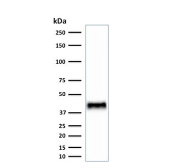
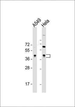
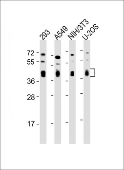
Western blot testing of mouse NIH3T3 cell lysate with BMI1 antibody. Predicted molecular weight: 37-43 kDa.

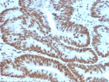
IHC staining of FFPE human colon carcinoma with BMI1 antibody. HIER: boil tissue sections in pH9 10mM Tris with 1mM EDTA for 20 min and allow to cool before testing.
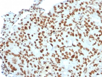
IHC staining of FFPE human breast carcinoma with BMI1 antibody. HIER: boil tissue sections in pH9 10mM Tris with 1mM EDTA for 20 min and allow to cool before testing.
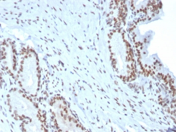
IHC staining of FFPE human prostate carcinoma with BMI1 antibody. HIER: boil tissue sections in pH9 10mM Tris with 1mM EDTA for 20 min and allow to cool before testing.

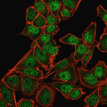
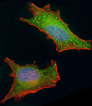
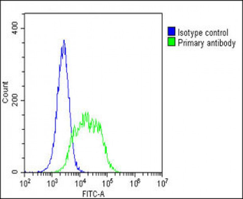
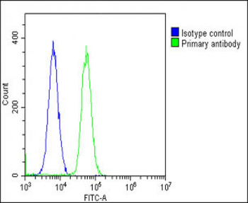
Immunofluorescent staining of PFA-fixed human HeLa cells with BMI1 antibody (green) and Phalloidin (red).

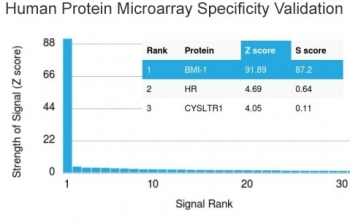
Analysis of HuProt (TM) microarray containing more than 19000 full-length human proteins using BMI1 antibody (clone BMI1/2823). These results demonstrate the foremost specificity of the BMI1/2823 mAb. Z- and S- score: The Z-score represents the strength of a signal that an antibody (in combination with a fluorescently-tagged anti-IgG secondary Ab) produces when binding to a particular protein on the HuProt (TM) array. Z-scores are described in units of standard deviations (SD's) above the mean value of all signals generated on that array. If the targets on the HuProt (TM) are arranged in descending order of the Z-score, the S-score is the difference (also in units of SD's) between the Z-scores. The S-score therefore represents the relative target specificity of an Ab to its intended target.
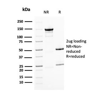
SDS-PAGE analysis of purified, BSA-free BMI1 antibody as confirmation of integrity and purity.
- Item 1 of 8
BMI1 antibody [orb19786]
ICC, IF, IHC-P, WB
Guinea pig, Human, Mouse, Rat
Rabbit
Polyclonal
Unconjugated
100 μg - Item 1 of 7
- Item 1 of 7
- Item 1 of 7
- Item 1 of 4
Goat anti-BMI1 (aa237-251) Antibody [orb233612]
ELISA, IHC, WB
Canine, Human, Mouse, Porcine
Goat
Polyclonal
Unconjugated
100 μg