You have no items in your shopping cart.
Cart summary
Item 1 of 7
Item 1 of 7
BMI1 Antibody
Catalog Number: orb1928300

| Catalog Number | orb1928300 |
|---|---|
| Category | Antibodies |
| Description | Affinity Purified Rabbit Polyclonal Antibody (Pab) |
| Species/Host | Rabbit |
| Clonality | Polyclonal |
| Clone Number | RB22858 |
| Tested applications | FC, IF, IHC-P, WB |
| Reactivity | Human, Mouse |
| Isotype | Rabbit IgG |
| Antibody Type | Primary Antibody |
| Dilution range | IF: 1:25, WB: 1:2000, WB: 1:8000, IHC-P: 1:10~50, FC: 1:25, FC: 1:25, FC: 1:25 |
| Form/Appearance | Purified polyclonal antibody supplied in PBS with 0.09% (W/V) sodium azide. This antibody is purified through a protein A column, followed by peptide affinity purification. |
| Conjugation | Unconjugated |
| MW | 36949 Da |
| Target | This BMI1 antibody is generated from rabbits immunized with BMI1 recombinant protein. |
| UniProt ID | P35226 |
| NCBI | NP_001190991.1, NP_005171.4 |
| Storage | Maintain refrigerated at 2-8°C for up to 2 weeks. For long term storage store at -20°C in small aliquots to prevent freeze-thaw cycles |
| Alternative names | Polycomb complex protein BMI-1, Polycomb group RIN Read more... |
| Note | For research use only |
| Expiration Date | 12 months from date of receipt. |

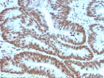
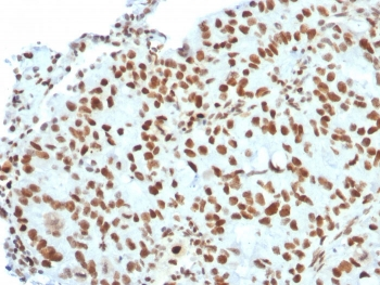
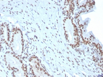
Formalin-fixed and paraffin-embedded human breast carcinoma reacted with BMI1 Antibody, which was peroxidase-conjugated to the secondary antibody, followed by DAB staining. This data demonstrates the use of this antibody for immunohistochemistry; clinical relevance has not been evaluated.
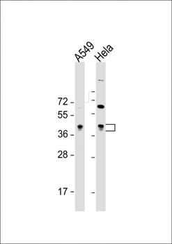
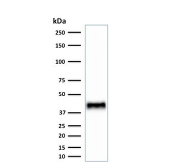
All lanes: Anti-BMI1 Antibody at 1:8000 dilution. Lane 1: A549 whole cell lysate. Lane 2: Hela whole cell lysate. Lysates/proteins at 20 µg per lane. Secondary Goat Anti-Rabbit IgG, (H+L), Peroxidase conjugated at 1/10000 dilution. Predicted band size: 37 kDa. Blocking/Dilution buffer: 5% NFDM/TBST.
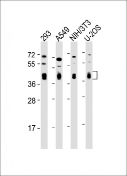
All lanes: Anti-BMI1 Antibody at 1:2000 dilution. Lane 1: 293 whole cell lysate. Lane 2: A549 whole cell lysate. Lane 3: NIH/3T3 whole cell lysate. Lane 4: U-2OS whole cell lysate. Lysates/proteins at 20 µg per lane. Secondary Goat Anti-Rabbit IgG, (H+L), Peroxidase conjugated at 1/10000 dilution. Predicted band size: 37 kDa. Blocking/Dilution buffer: 5% NFDM/TBST.
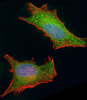
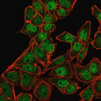
Immunofluorescent analysis of 4% paraformaldehyde-fixed, 0.1% Triton X-100 permeabilized HeLa (human cervical epithelial adenocarcinoma cell line) cells labeling BMI1 at 1/25 dilution, followed by Dylight 488-conjugated goat anti-rabbit IgG (1583138) secondary antibody at 1/200 dilution (green). Immunofluorescence image showing cytoplasm and nucleus staining on HeLa cell line. Cytoplasmic actin is detected with Dylight 554 Phalloidin at 1/100 dilution (red). The nuclear counter stain is DAPI (blue).
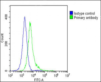
Overlay histogram showing Hela cells stained (green line). The cells were fixed with 2% paraformaldehyde (10 min) and then permeabilized with 90% methanol for 10 min. The cells were then icubated in 2% bovine serum albumin to block non-specific protein-protein interactions followed by the antibody (1:25 dilution) for 60 min at 37°C. The secondary antibody used was Goat-Anti-Rabbit IgG, DyLight 488 Conjugated Highly Cross-Adsorbed at 1/200 dilution for 40 min at 37°C. Isotype control antibody (blue line) was rabbit IgG1 (1 μg/1x10^6 cells) used under the same conditions. Acquisition of > 10000 events was performed.
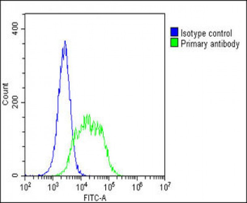
Overlay histogram showing A549 cells stained (green line). The cells were fixed with 2% paraformaldehyde (10 min) and then permeabilized with 90% methanol for 10 min. The cells were then icubated in 2% bovine serum albumin to block non-specific protein-protein interactions followed by the antibody (1:25 dilution) for 60 min at 37°C. The secondary antibody used was Goat-Anti-Rabbit IgG, DyLight 488 Conjugated Highly Cross-Adsorbed at 1/200 dilution for 40 min at 37°C. Isotype control antibody (blue line) was rabbit IgG1 (1 μg/1x10^6 cells) used under the same conditions. Acquisition of > 10000 events was performed.
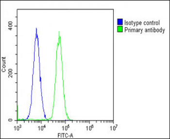
Overlay histogram showing U-2 OS cells stained (green line). The cells were fixed with 2% paraformaldehyde (10 min) and then permeabilized with 90% methanol for 10 min. The cells were then icubated in 2% bovine serum albumin to block non-specific protein-protein interactions followed by the antibody (1:25 dilution) for 60 min at 37°C. The secondary antibody used was Goat-Anti-Rabbit IgG, DyLight 488 Conjugated Highly Cross-Adsorbed at 1/200 dilution for 40 min at 37°C. Isotype control antibody (blue line) was rabbit IgG1 (1 μg/1x10^6 cells) used under the same conditions. Acquisition of > 10000 events was performed.
- Item 1 of 8
BMI1 antibody [orb19786]
ICC, IF, IHC-P, WB
Guinea pig, Human, Mouse, Rat
Rabbit
Polyclonal
Unconjugated
100 μg - Item 1 of 7
- Item 1 of 7
- Item 1 of 7
- Item 1 of 4
Goat anti-BMI1 (aa237-251) Antibody [orb233612]
ELISA, IHC, WB
Canine, Human, Mouse, Porcine
Goat
Polyclonal
Unconjugated
100 μg