You have no items in your shopping cart.
Cart summary
Item 1 of 3
Item 1 of 3
BIN1 antibody
Catalog Number: orb344474

| Catalog Number | orb344474 |
|---|---|
| Category | Antibodies |
| Description | BIN1 antibody |
| Species/Host | Mouse |
| Clonality | Monoclonal |
| Clone Number | 99F |
| Tested applications | ELISA, IF, IP, WB |
| Reactivity | Human, Mouse |
| Isotype | IgG |
| Immunogen | Anti-BIN1 (MOUSE) Monoclonal Antibody was produced in mouse by repeated immunizations with BIN1 polypeptide followed by hybridoma development. |
| Concentration | 1 mg/ml |
| Dilution range | ELISA: 1:5000 - 1:50000, IF: 1:100-1:500, IP: 10-100 µL, WB: 1:500-1:1500 |
| Form/Appearance | Liquid (sterile filtered) |
| Purity | Anti-BIN1 was purified from concentrated tissue culture supernate by Protein G chromatography followed by extensive dialysis against the buffer stated above. BIN1 antibody is specific for human BIN1 protein. A BLAST analysis was used to suggest cross-reactivity with BIN1 from human and mouse sources based on 100% homology with the immunizing sequence. Cross-reactivity with BIN1 from other sources has not been determined. |
| Conjugation | Unconjugated |
| UniProt ID | O00499 |
| NCBI | NP_004296.1 |
| Storage | Store vial at -20° C or below prior to opening. This vial contains a relatively low volume of reagent (25 µL). To minimize loss of volume dilute 1:10 by adding 225 µL of the buffer stated above directly to the vial. Recap, mix thoroughly and briefly centrifuge to collect the volume at the bottom of the vial. Use this intermediate dilution when calculating final dilutions as recommended below. Store the vial at -20°C or below after dilution. Avoid cycles of freezing and thawing. |
| Buffer/Preservatives | 0.01% (w/v) Sodium Azide |
| Alternative names | mouse anti-BIN1 Antibody, AMPHL, Myc box-dependent Read more... |
| Note | For research use only |
| Application notes | Anti-BIN1 antibody has been tested for use in ELISA, Western Blot, IP, and IF. Specific conditions for reactivity should be optimized by the end user. |
| Expiration Date | 12 months from date of receipt. |
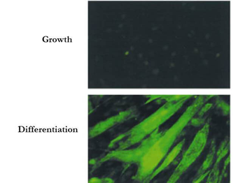
Immunofluorescence Microscopy of Mouse Anti-BIN1 Antibody. Cells: C2C12 cells during growth or differentiation. Fixation: 0.5% PFA. Antigen retrieval: not required. Primary antibody: BIN-1 (Exon 10 specific, 99F) monoclonal antibody. Secondary antibody: mouse secondary antibody at 1:10000 for 45 min at RT. Localization: BIN1 is nuclear and cytoplasmic. Staining: BIN 1 as green fluorescent signal.
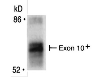
Immunoprecipitation of Mouse anti-BIN1 antibody. Immunoprecipitates were separated by SDS-PAGE and visualized by fluorography. The differentiation-associated (Exon 10+) form of Bin1 is recognized by using Bin1 (Exon 10 specific, 99F) monoclonal antibody. Lane 1: BIN1 [35 µg]. Primary antibody: Anti-BIN1 (Exon 10 specific, 99F) monoclonal antibody at 1:400 for overnight at 2-8°C. Secondary antibody: IRDye800™ mouse secondary antibody at 1:10000 for 45 min at RT. Block: 5% BLOTTO overnight at 4°C. Predicted MW: 64.7 kDa. Observed MW: ~65 kDa for BIN-1.
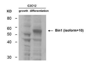
Western Blot of Mouse Anti-BIN1 antibody. Lane 1: C2C12 during growth. Lane 2: C2C12 during differentiation. Load: 35 µg per lane. Primary antibody: BIN1 antibody at 1:400 for overnight at 4°C. Secondary antibody: IRDye800™ mouse secondary antibody at 1:10000 for 45 min at RT. Block: 5% BLOTTO overnight at 4°C. Predicted/Observed size: 64.7 kDa, ~55 kDa for BIN-1. Other band(s): non-specifics.
- Item 1 of 6
- Item 1 of 6
BIN1 Antibody [orb865488]
ELISA, FC, ICC, IF, IHC, WB
Human, Mouse, Rat
Rabbit
Polyclonal
Unconjugated
10 μg, 100 μg - Item 1 of 5
- Item 1 of 5
- Item 1 of 4


Submit a review
Filter by Rating

- 5 stars

- 4 stars
- 3 stars
- 2 stars
- 1 stars
