You have no items in your shopping cart.
Cart summary
Item 1 of 3
Item 1 of 3
BIN1 antibody
Catalog Number: orb344473

| Catalog Number | orb344473 |
|---|---|
| Category | Antibodies |
| Description | BIN1 antibody |
| Species/Host | Mouse |
| Clonality | Monoclonal |
| Clone Number | 99F |
| Tested applications | ELISA, IF, IP, WB |
| Reactivity | Human, Mouse |
| Isotype | IgG |
| Immunogen | Anti-BIN1 (MOUSE) Monoclonal Antibody was produced in mouse by repeated immunizations with BIN1 polypeptide followed by hybridoma development. |
| Concentration | 1 mg/ml |
| Dilution range | ELISA: 1:5000 - 1:50000, IF: 1:100-1:500, IP: 10-100 µL, WB: 1:500-1:1500 |
| Form/Appearance | Liquid (sterile filtered) |
| Purity | Anti-BIN1 was purified from concentrated tissue culture supernate by Protein G chromatography followed by extensive dialysis against the buffer stated above. BIN1 antibody is specific for human BIN1 protein. A BLAST analysis was used to suggest cross-reactivity with BIN1 from human and mouse sources based on 100% homology with the immunizing sequence. Cross-reactivity with BIN1 from other sources has not been determined. |
| Conjugation | Unconjugated |
| UniProt ID | O00499 |
| NCBI | NP_004296.1 |
| Storage | Store vial at -20° C prior to opening. Aliquot contents and freeze at -20° C or below for extended storage. Avoid cycles of freezing and thawing. Centrifuge product if not completely clear after standing at room temperature. This product is stable for several weeks at 4° C as an undiluted liquid. Dilute only prior to immediate use. |
| Buffer/Preservatives | 0.01% (w/v) Sodium Azide |
| Alternative names | mouse anti-BIN1 Antibody, AMPHL, Myc box-dependent Read more... |
| Note | For research use only |
| Application notes | Anti-BIN1 antibody has been tested for use in ELISA, Western Blot, IP, and IF. Specific conditions for reactivity should be optimized by the end user. |
| Expiration Date | 12 months from date of receipt. |
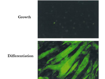
Immunofluorescence Microscopy of Mouse Anti-BIN1 Antibody. Cells: C2C12 cells during growth or differentiation. Fixation: 0.5% PFA. Antigen retrieval: not required. Primary antibody: BIN-1 (Exon 10 specific, 99F) monoclonal antibody. Secondary antibody: mouse secondary antibody at 1:10000 for 45 min at RT. Localization: BIN1 is nuclear and cytoplasmic. Staining: BIN 1 as green fluorescent signal.
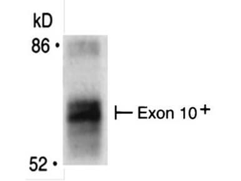
Immunoprecipitation of Mouse anti-BIN1 antibody. Immunoprecipitates were separated by SDS-PAGE and visualized by fluorography. The differentiation-associated (Exon 10+) form of Bin1 is recognized by using Bin1 (Exon 10 specific, 99F) monoclonal antibody. Lane 1: BIN1 [35 µg]. Primary antibody: Anti-BIN1 (Exon 10 specific, 99F) monoclonal antibody at 1:400 for overnight at 2-8°C. Secondary antibody: IRDye800™ mouse secondary antibody at 1:10000 for 45 min at RT. Block: 5% BLOTTO overnight at 4°C. Predicted MW: 64.7 kDa. Observed MW: ~65 kDa for BIN-1.
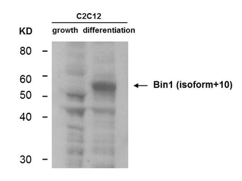
Western Blot of Mouse Anti-BIN1 antibody. Lane 1: C2C12 during growth. Lane 2: C2C12 during differentiation. Load: 35 µg per lane. Primary antibody: BIN1 antibody at 1:400 for overnight at 4°C. Secondary antibody: IRDye800™ mouse secondary antibody at 1:10000 for 45 min at RT. Block: 5% BLOTTO overnight at 4°C. Predicted/Observed size: 64.7 kDa, ~55 kDa for BIN-1. Other band(s): non-specifics.
- Item 1 of 6
- Item 1 of 6
BIN1 Antibody [orb865488]
ELISA, FC, ICC, IF, IHC, WB
Human, Mouse, Rat
Rabbit
Polyclonal
Unconjugated
10 μg, 100 μg - Item 1 of 5
- Item 1 of 5
- Item 1 of 4


Submit a review
Filter by Rating

- 5 stars

- 4 stars
- 3 stars
- 2 stars
- 1 stars
