You have no items in your shopping cart.
Cart summary
Item 1 of 3
Item 1 of 3
Beta galactosidase antibody (FITC)
Catalog Number: orb344848

| Catalog Number | orb344848 |
|---|---|
| Category | Antibodies |
| Description | Beta galactosidase antibody (FITC) |
| Species/Host | Rabbit |
| Clonality | Polyclonal |
| Tested applications | IF, WB |
| Isotype | IgG |
| Immunogen | Beta Galactosidase (E. coli) |
| Concentration | 5.0 mg/mL |
| Dilution range | IF: 1:500 - 1:2,500, WB: 1:10,000 |
| Form/Appearance | Lyophilized |
| Purity | This product is an IgG fraction antibody purified from monospecific antiserum by a multi-step process which includes delipidation, salt fractionation and ion exchange chromatography followed by extensive dialysis against the buffer stated above. Assay by immunoelectrophoresis resulted in a single precipitin arc against anti-fluorescein, anti-Rabbit Serum and purified and partially purified Beta Galactosidase (E. coli). |
| Conjugation | FITC |
| UniProt ID | P00722 |
| NCBI | NP_414878.1 |
| Storage | Store vial antibody at 4° C prior to restoration. For extended storage aliquot contents and freeze at -20° C or below. Avoid cycles of freezing and thawing. Centrifuge product if not completely clear after standing at room temperature. This product is stable for several weeks at 4° C as an undiluted liquid. Dilute only prior to immediate use. |
| Buffer/Preservatives | 0.01% (w/v) Sodium Azide |
| Alternative names | rabbit anti-Beta Galactosidase Antibody fluorescei Read more... |
| Note | For research use only |
| Application notes | Anti-Beta Galactosidase Fluorescein Conjugated Antibody has been tested by western blot and is designed for fluorescent western blotting, also suitable for multiplex analysis, including multicolor imaging, utilizing various commercial platforms. |
| Expiration Date | 12 months from date of receipt. |

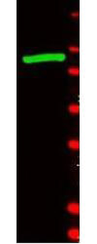
Western blot using Biorbyt's anti-b-Galactosidase antibody shows detection of a band at ~117 kDa (lane 1) corresponding to b-Gal present in a partially purified preparation (arrowhead). Approximately 1 µg of protein was resolved on a 4-20% Tris-Glycine gel by SDS-PAGE and transferred onto nitrocellulose. After blocking, the membrane was probed with the primary antibody diluted to 1:1000. Reaction occurred overnight at 4°C followed by washes and reaction with a 1:10000 dilution of IRDye® 800 conjugated Gt-a-Rabbit IgG (H&L) MX10 for 45 min at room temperature (800 nm channel, green). Molecular weight estimation was made by comparison to prestained MW markers in lane M (700 nm channel, red).
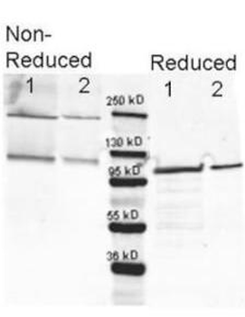
Western blotting using Biorbyt's anti-b-Galactosidase antibody. Lane 1 shows 80 ng and lane 2 shows 20 ng loaded onto gel. Results for non-reducing conditions of SDS-PAGE prior to transfer to nitrocellulose are shown on the left side of the figure; results obtainined under reducing conditions are shown on the right. Blots were blocked overnight at 4°C with Blocking Buffer for Fluorescent Western Blotting (p/n orb348637). The membrane was probed with anti-b-Galactosidase diluted to 1:10000. Reaction occurred overnight at 4°C. Dylight649™ conjugated Gt-a-anti-Rabbit IgG was used for detection. Molecular weight estimation was made by comparison to a prestained MW marker (center).in lane M.

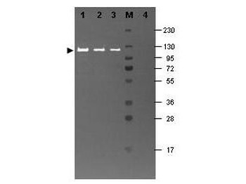
Western blotting using Biorbyt's Fluorescein conjugated anti-b-Galactosidase antibody shows a band at ~117 kDa (lanes 1 - 3) corresponding to 60 ng, 30 ng and 15 ng, respectively of b-Gal present in partially purified preparations (arrowhead). Lane 4 shows no cross reactivity with proteins present in a non-specific control E.coli lysate. Proteins were resolved on a 4-20% Tris-Glycine gel by SDS-PAGE and transferred to nitrocellulose and blocking using Blocking Buffer for Fluorescent Western Blotting (p/n orb348637). The membrane was probed with fluorescein conjugated anti-b-Galactosidase (p/n orb344848) diluted to 1:10000. Reaction occurred for 2 hours at room temperature. Molecular weight estimation was made by comparison to a prestained MW marker in lane M.
- Item 1 of 3
- Item 1 of 3


Submit a review
Filter by Rating

- 5 stars

- 4 stars
- 3 stars
- 2 stars
- 1 stars
