You have no items in your shopping cart.
Cart summary
Item 1 of 3
Item 1 of 3
Beta Actin Antibody / ACTB
Catalog Number: orb2634887

| Catalog Number | orb2634887 |
|---|---|
| Category | Antibodies |
| Description | All eukaryotic cells express Actin, which often constitutes as much as 50% of total cellular protein. Actin filaments can form both stable and labile structures and are crucial components of microvilli and the contractile apparatus of muscle cells. While lower eukaryotes, such as yeast, have only one Actin gene, higher eukaryotes have several isoforms encoded by a family of genes. At least six types of Actin are present in mammalian tissues and fall into three classes. a-Actin expression is limited to various types of muscle, whereas -Actin and g-Actin are the principle constituents of filaments in other tissues. Members of the small GTPase family regulate the organization of the Actin cytoskeleton. Rho controls the assembly of Actin stress fibers and focal adhesion. Rac regulates Actin filament accumulation at the plasma membrane. Cdc42 stimulates formation of filopodia. |
| Species/Host | Mouse |
| Clonality | Monoclonal |
| Clone Number | ACTB/1108 |
| Tested applications | IHC-P, WB |
| Reactivity | Human, Mouse, Rat |
| Isotype | Mouse IgG2b, kappa |
| Immunogen | Recombinant human ACTB protein was used as the immunogen for the Beta Actin antibody. |
| Dilution range | Western blot: 1-2ug/ml,Immunohistochemistry (FFPE): 1-2ug/ml |
| Conjugation | Unconjugated |
| Formula | 0.2 mg/ml in 1X PBS with 0.1 mg/ml BSA (US sourced), 0.05% sodium azide |
| Hazard Information | This Beta Actin antibody is available for research use only. |
| UniProt ID | P60709 |
| Storage | Maintain refrigerated at 2-8°C for up to 2 weeks. For long term storage store at -20°C in small aliquots to prevent freeze-thaw cycles. |
| Note | For research use only |
| Application notes | Optimal dilution of the Beta Actin antibody should be determined by the researcher. |
| Expiration Date | 12 months from date of receipt. |

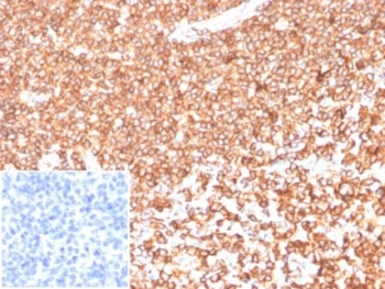
IHC staining of FFPE human thyroid tissue with Beta Actin (clone ACTB/1108). Negative control inset: PBS instead of primary antibody to control for secondary binding. HIER: boil tissue sections in pH9 10mM Tris with 1mM EDTA for 20 min and allow to cool before testing.

IHC staining of FFPE human tonsil tissue with Beta Actin antibody (clone ACTB/1108). Negative control inset: PBS used instead of primary antibody to control for secondary Ab binding. HIER: boil tissue sections in pH9 10mM Tris with 1mM EDTA for 20 min and allow to cool before testing.
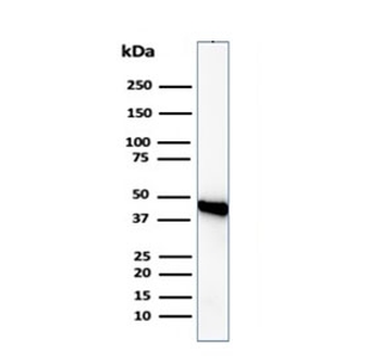
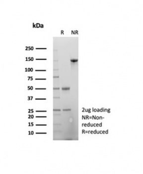
Western blot analysis of human Raji cell lysate using Beta Actin antibody (clone ACTB/1108). Predicted molecular weight ~42 kDa.
- Item 1 of 3
Beta Actin Antibody / ACTB [orb2634886]
IHC-P, WB
Human, Mouse, Rat
Mouse
Monoclonal
Unconjugated
100 μg - Item 1 of 3
Beta Actin Antibody / ACTB [orb2634888]
IHC-P, WB
Human, Mouse, Rat
Mouse
Monoclonal
Unconjugated
100 μg - Item 1 of 2
- Item 1 of 2
- Item 1 of 2