You have no items in your shopping cart.
Cart summary
Item 1 of 13
Item 1 of 13
Beclin-1 Antibody
Catalog Number: orb1239257

| Catalog Number | orb1239257 |
|---|---|
| Category | Antibodies |
| Description | Beclin-1 Antibody |
| Species/Host | Rabbit |
| Clonality | Polyclonal |
| Tested applications | ELISA, IF, IHC-P, WB |
| Predicted Reactivity | Bovine, Porcine |
| Reactivity | Human, Mouse, Rat |
| Isotype | IgG |
| Immunogen | Anti-Beclin-1 antibody (orb1239257) was raised against a peptide corresponding to 17 amino acids near the center of human Beclin-1. The immunogen is located between 40-90 amino acids of Beclin-1. |
| Concentration | 1 mg/mL |
| Form/Appearance | Liquid |
| Conjugation | Unconjugated |
| MW | Predicted: 52 kDObserved: 60 kD |
| Target | BECN1 |
| UniProt ID | Q14457 |
| NCBI | AAH10276 |
| Storage | Maintain refrigerated at 2-8°C for up to 2 weeks. For long term storage store at -20°C in small aliquots to prevent freeze-thaw cycles. |
| Buffer/Preservatives | Beclin-1 Antibody is supplied in PBS containing 0.02% sodium azide. |
| Alternative names | Beclin-1 Antibody: ATG6, VPS30, beclin1, GT197, Be Read more... |
| Note | For research use only |
| Expiration Date | 12 months from date of receipt. |

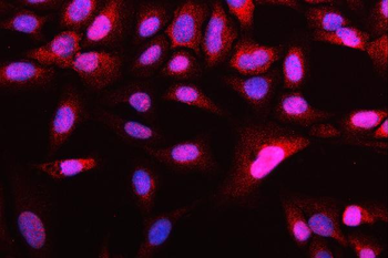
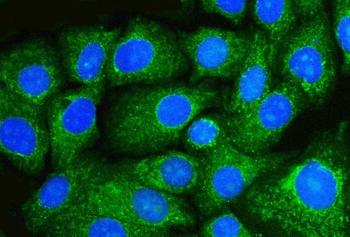
Immunofluorescence Validation of Beclin-1 in HeLa Cells. Immunofluorescent analysis of methanol-fixed HeLa cells labeling Beclin-1 with orb1239257 at 10 µg/mL, followed by goat anti-rabbit IgG secondary antibody at 1/1000 dilution (red) and DAPI staining (blue). Alpha tubulin was stained with anti-alpha tubulin antibody following by goat anti-mouse IgG secondary antibody (green). Images were captured with confocal microscopy.
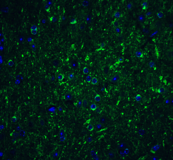
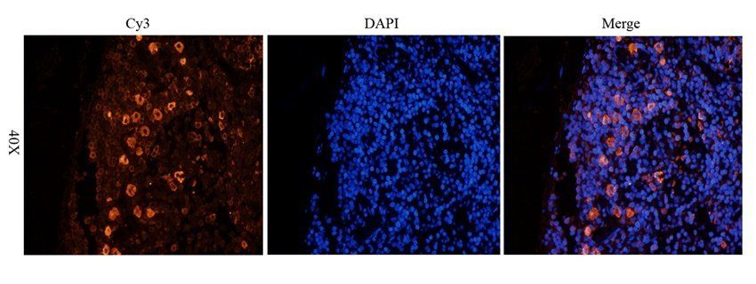
Immunofluorescence Validation of Beclin-1 in Mouse Brain Tissue. Immunofluorescent analysis of 4% paraformaldehyde-fixed mouse brain labeling Beclin-1 with orb1239257 at 20 g/mL, followed by goat anti-rabbit IgG secondary antibody at 1/500 dilution (green) and DAPI antibody (blue).
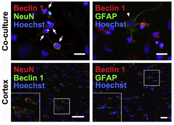
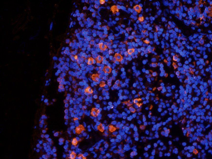
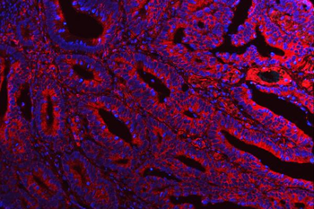
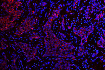
Immunofluorescent Validation of Beclin-1 in Human Cortical Neurons and Rat Brain. Cell specificity of Beclin 1 expression. Cortical neurons and glia co-cultures (upper panels) and cortex slices (lower panels) were immunostained with anti-Beclin 1 (red) and NeuN (neuronal biomarker, green) or GFAP (astrocyte biomarker, green). Neurons and astrocyte in cultured cells were indicated by arrows and arrowhead, respectively. Areas in white boxes were enlarged.
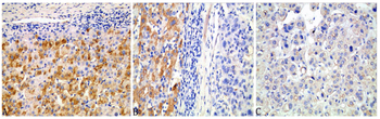
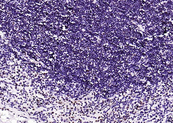
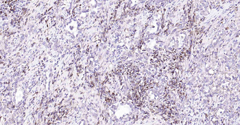
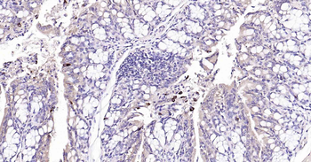
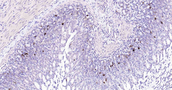
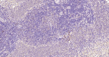
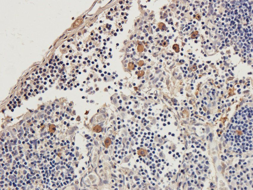
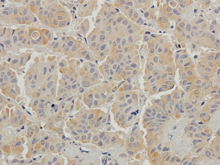
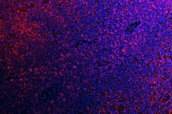
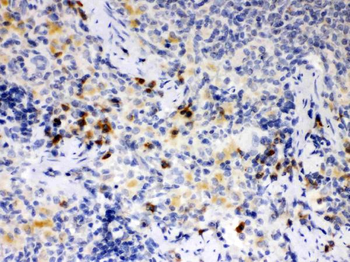
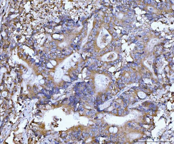
Immunohistochemistry Validation of Beclin-1 in Human HCC Cells. Beclin-1 exhibited cytoplasmic staining in adjacent non-tumor (ANT) tissues (A), bordeling site between HCC and ANT (B: left, ANT; right: HCC), and HCC (C), respectively. Beclin-1 expression was stronger in ANT than in HCC. Magnification × 400.
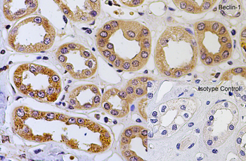
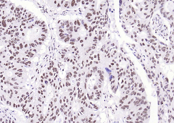
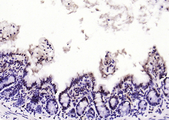
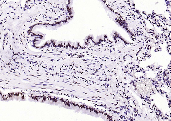
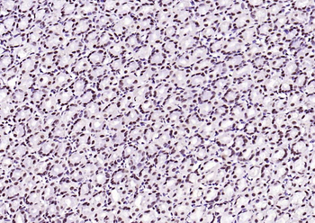
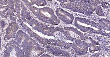
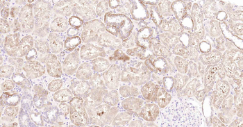
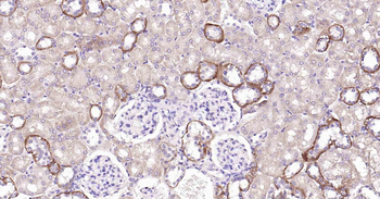
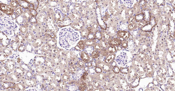
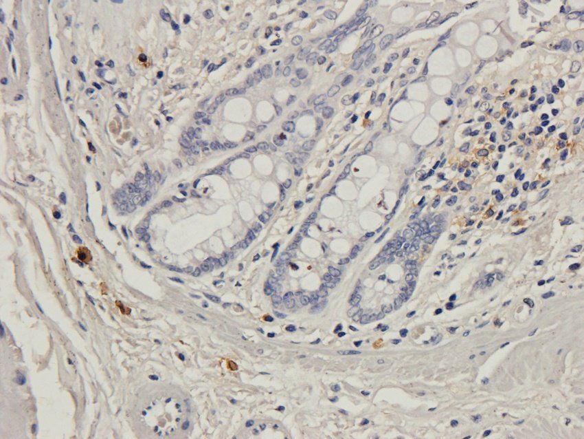
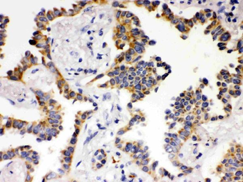
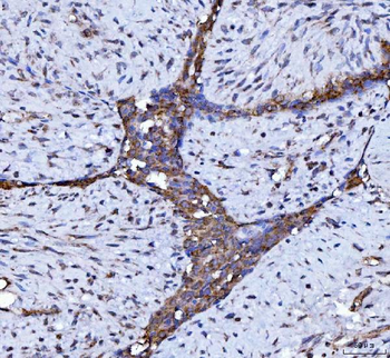
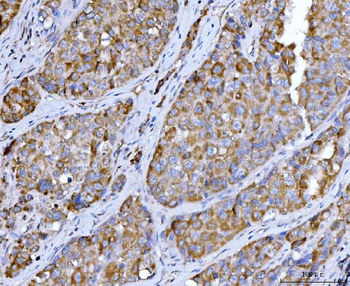
Immunohistochemistry Validation of Beclin-1 in Human kidney Tissue. Immunohistochemical analysis of paraffin-embedded human kidney tissue using anti-Beclin-1 antibody (orb1239257) at 2 µg/mL. Tissue was fixed with formaldehyde and blocked with 10% serum for 1 h at RT; antigen retrieval was by heat mediation with a citrate buffer (pH6). Samples were incubated with primary antibody overnight at 4°C. A goat anti-rabbit IgG H&L (HRP) at 1/250 was used as secondary. Counter stained with Hematoxylin.
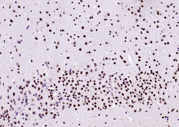
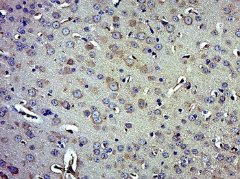
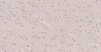
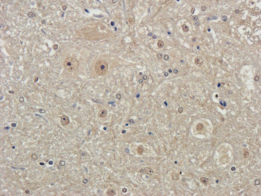
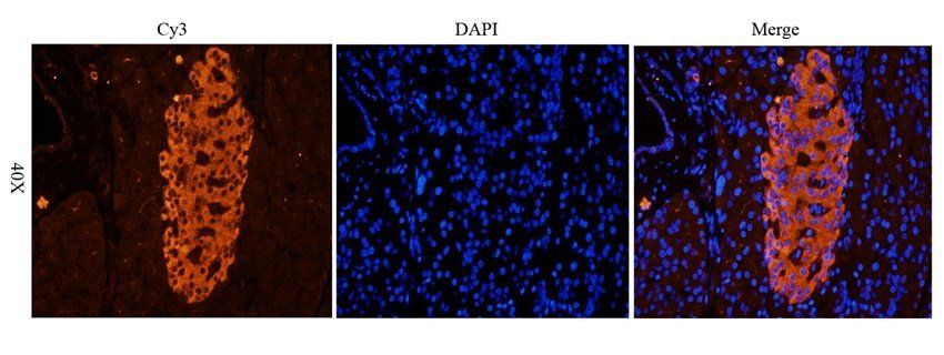
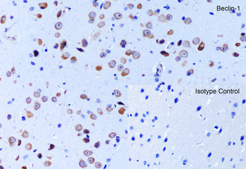
Immunohistochemistry Validation of Beclin-1 in Mouse Brain Tissue. Immunohistochemical analysis of paraffin-embedded mouse brain tissue using anti-Beclin-1 antibody (orb1239257) at 5 µg/mL. Tissue was fixed with formaldehyde and blocked with 10% serum for 1 h at RT; antigen retrieval was by heat mediation with a citrate buffer (pH6). Samples were incubated with primary antibody overnight at 4°C. A goat anti-rabbit IgG H&L (HRP) at 1/250 was used as secondary. Counter stained with Hematoxylin.
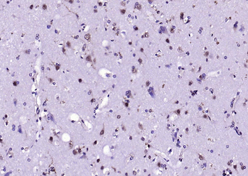
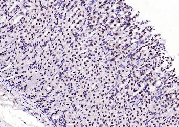
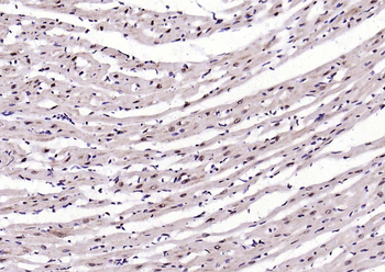
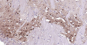
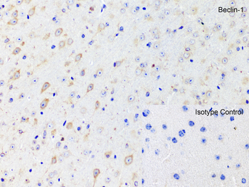
Immunohistochemistry Validation of Beclin-1 in Rat Brain Tissue. Immunohistochemical analysis of paraffin-embedded rat brain tissue using anti-Beclin-1 antibody (orb1239257) at 2 µg/mL. Tissue was fixed with formaldehyde and blocked with 10% serum for 1 h at RT; antigen retrieval was by heat mediation with a citrate buffer (pH6). Samples were incubated with primary antibody overnight at 4°C. A goat anti-rabbit IgG H&L (HRP) at 1/250 was used as secondary. Counter stained with Hematoxylin.
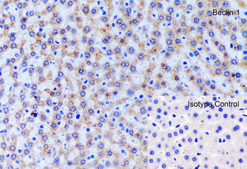
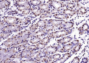
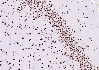
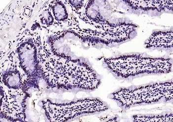
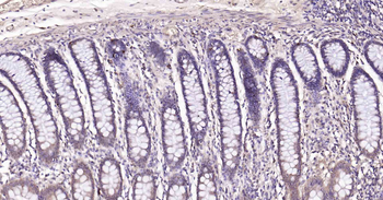
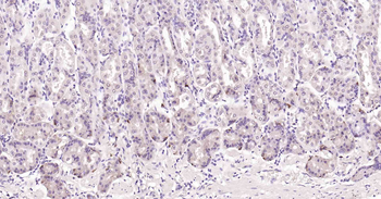
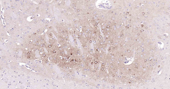
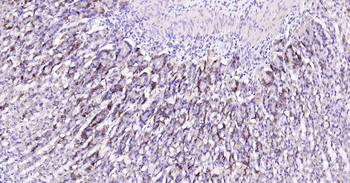
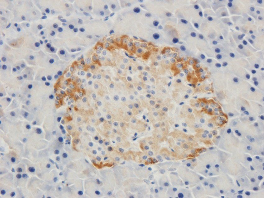
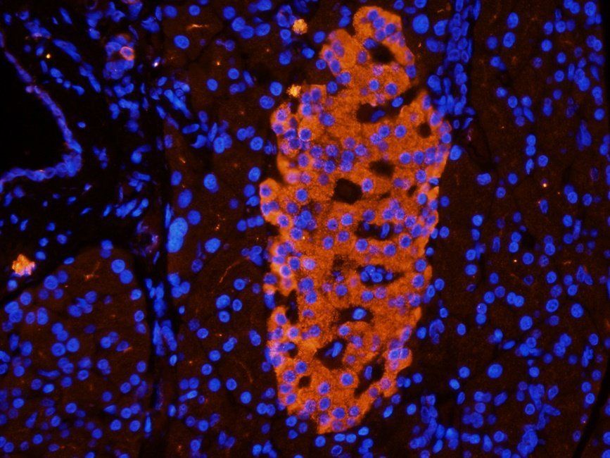
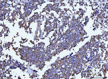
Immunohistochemistry Validation of Beclin-1 in Rat Liver Tissue. Immunohistochemical analysis of paraffin-embedded rat liver tissue using anti-Beclin-1 antibody (orb1239257) at 2 µg/mL. Tissue was fixed with formaldehyde and blocked with 10% serum for 1 h at RT; antigen retrieval was by heat mediation with a citrate buffer (pH6). Samples were incubated with primary antibody overnight at 4°C. A goat anti-rabbit IgG H&L (HRP) at 1/250 was used as secondary. Counter stained with Hematoxylin.
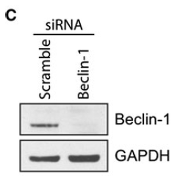
Knockdown of Beclin-1 in Human T47D Cells. T47D cells were transfected with scrambled or Beclin-1 siRNA. The following day, these cells were treated with vehicle, C, 0.5 mM VPA, 10 lM 4OH-tamoxifen, T, or 0.5 mM VPA and 10 lM 4OH-tamoxifen, VT, for 48 h and viability was assayed by dye exclusion.
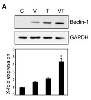
Regulated Expression of Beclin-1 in Human T47D Cells. Treatment with tamoxifen and an HDAC inhibitor alters the balance of autophagy and apoptosis inhibitors and drivers. T47D cells were treated with vehicle, C, 0.5 mM VPA, V, 10 lM 4OHtamoxifen, T, or 0.5 mM VPA and 10 lM 4OH-tamoxifen, VT, for 48 h and Beclin-1 protein and RNA.
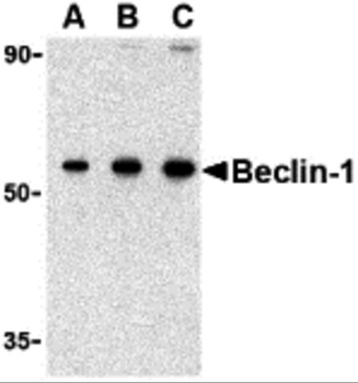
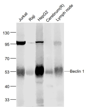
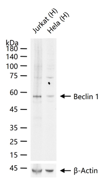
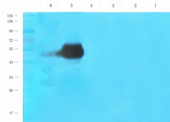
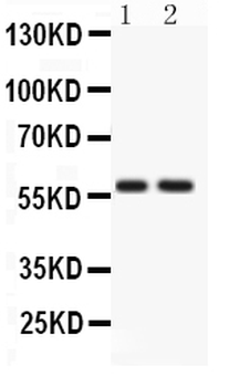
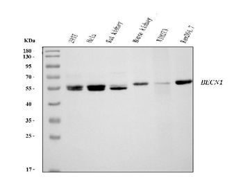
WB Validation in Human Cell Lines. Loading: 15 µg of lysate Antibodies: Beclin-1 orb1239257, 1 µg/mL, 1 h incubation at RT in 5% NFDM/TBST. Secondary: Goat Anti-Rabbit IgG HRP conjugate at 1:10000 dilution.
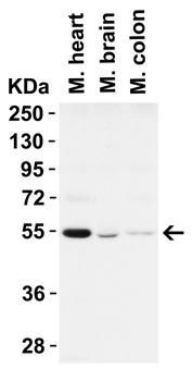
WB Validation in Mouse Tissues. Loading: 15 µg of lysate Antibodies: Beclin-1 orb1239257, 1 µg/mL, 1 h incubation at RT in 5% NFDM/TBST. Secondary: Goat Anti-Rabbit IgG HRP conjugate at 1:10000 dilution.
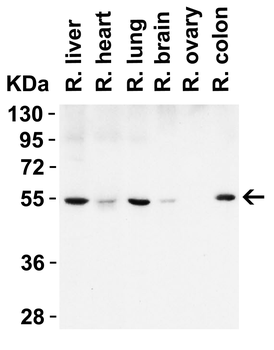
WB Validation in Rat Tissues. Loading: 15 µg of lysate Antibodies: Beclin-1 orb1239257, 1 µg/mL, 1 h incubation at RT in 5% NFDM/TBST. Secondary: Goat Anti-Rabbit IgG HRP conjugate at 1:10000 dilution.
- Item 1 of 16
Beclin 1 Mouse Monoclonal Antibody [orb499660]
IF, IHC-Fr, IHC-P
Mouse, Rat
Human, Mouse, Rat
Mouse
Monoclonal
Unconjugated
200 μl, 100 μl, 50 μl - Item 1 of 18
Beclin 1 Recombinant Rabbit Monoclonal Antibody [orb2563484]
IF, IHC-Fr, IHC-P, WB
Mouse, Rat
Human, Mouse, Rat
Rabbit
Recombinant
Unconjugated
200 μl, 50 μl, 100 μl - Item 1 of 10
- Item 1 of 10
Anti-ATG14L Antibody [orb308776]
FC, ICC, IF, IHC, WB
Human, Rat
Rabbit
Polyclonal
Unconjugated
10 μg, 100 μg - Item 1 of 7
Anti-Beclin 1 Antibody (monoclonal, 2D12A3) [orb1145791]
FC, ICC, IF, IHC, WB
Human, Mouse, Rat
Mouse
Monoclonal
Unconjugated
10 μg, 100 μg