You have no items in your shopping cart.
| Catalog Number | orb4655 |
|---|---|
| Category | Antibodies |
| Description | Bax Rabbit Polyclonal Antibody |
| Species/Host | Rabbit |
| Clonality | Polyclonal |
| Tested applications | FC, ICC, IF, IHC-Fr, IHC-P, WB |
| Predicted Reactivity | Bovine, Canine, Porcine, Sheep |
| Reactivity | Human, Mouse, Rabbit, Rat |
| Isotype | IgG |
| Immunogen | KLH conjugated synthetic peptide derived from human Bax (84-175/192aa) |
| Concentration | 1mg/ml |
| Dilution range | WB=1:500-2000, IHC-P=1:100-500, IHC-F=1:100-500, ICC/IF=1:100, IF=1:100-500, Flow-Cyt=1μg /test |
| Form/Appearance | Liquid |
| Conjugation | Unconjugated |
| MW | 21 kDa |
| Target | BAX |
| UniProt ID | Q07812 |
| RRID | AB_10919579 |
| Storage | Maintain refrigerated at 2-8°C for up to 2 weeks. For long term storage store at -20°C in small aliquots to prevent freeze-thaw cycles. |
| Buffer/Preservatives | 0.01M TBS (pH7.4) with 1% rAlbumin, 0.02% Proclin300 and 50% Glycerol. |
| Alternative names | apoptosis regulator BAX; Apoptosis regulator BAX c Read more... |
| Note | For research use only |
| Expiration Date | 12 months from date of receipt. |
Mahdy, Heba M. et al. The anti-apoptotic and anti-inflammatory properties of puerarin attenuate 3-nitropropionic-acid induced neurotoxicity in rats Can J Physiol Pharmacol, 92, 252-258 (2014)
Guan, Jianmin et al. Long non‑coding RNA ZEB2‑AS1 affects cell proliferation and apoptosis via the miR‑122‑5p/PLK1 axis in acute myeloid leukemia Int. J. Mol. Med., (2020)
Mantawy, Eman M. et al. Chrysin alleviates acute doxorubicin cardiotoxicity in rats via suppression of oxidative stress, inflammation and apoptosis Eur J Pharmacol, 728, 107-118 (2014)
Ibrahim, Kamilia M. et al. Protection from doxorubicin-induced nephrotoxicity by clindamycin: novel antioxidant, anti-inflammatory and anti-apoptotic roles Naunyn Schmiedebergs Arch Pharmacol, 393, 739-748 (2020)
Herbstein, A. U. Limbal incision with conjunctival continuous key-pattern suture in squint surgery Br J Ophthalmol, 56, 703-705 (1972)
Carlisle, H. N. et al. Therapy of staphylococcal infections in monkeys. VI. Comparison of clindamycin, erythromycin, and methicillin Appl Microbiol, 21, 440-446 (1971)

Sample: Hela (Human) Cell Lysate at 30 ug, Primary: Anti-Bax (orb4655) at 1/1000 dilution, Secondary: IRDye800CW Goat Anti-Rabbit IgG at 1/20000 dilution, Predicted band size: 21 kD, Observed band size: 23 kD.
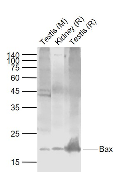
Sample: Lane 1: Testis (Mouse) Lysate at 40 ug, Lane 2: Kidney (Rat) Lysate at 40 ug, Lane 3: Testis (Rat) Lysate at 40 ug, Primary: Anti-Bax (orb4655) at 1/1000 dilution, Secondary: IRDye800CW Goat Anti-Rabbit IgG at 1/20000 dilution, Predicted band size: 21 kD, Observed band size: 21 kD.
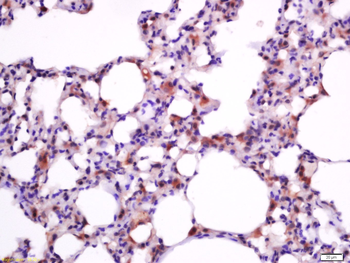
Tissue/Cell: rat lung tissue, 4% Paraformaldehyde-fixed and paraffin-embedded, Antigen retrieval: citrate buffer (0.01M, pH 6.0), Boiling bathing for 15 min, Block endogenous peroxidase by 3% Hydrogen peroxide for 30 min, Blocking buffer (normal goat serum) at 37°C for 20 min, Incubation: Anti-Bax Polyclonal Antibody, Unconjugated (orb4655) 1:200, overnight at 4°C, followed by conjugation to the secondary antibody and DAB staining.
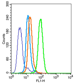
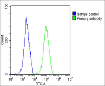
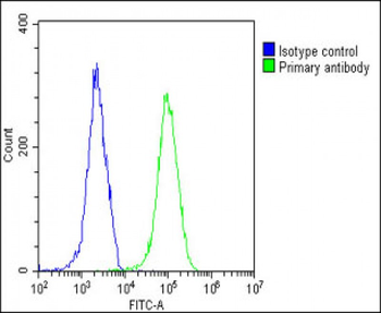
Overlay histogram showing HL 60 cells stained with orb4655 (Green line). The cells were fixed with 90% methanol (5 min) and then permeabilized with 0.01M PBS-Tween for 20 min. The cells were then incubated in 1x PBS / 10% normal goat serum to block non-specific protein-protein interactions followed by the antibody (orb4655, 1 µg/1x10^6 cells) for 30 min at 22°C. The secondary antibody used was fluorescein isothiocyanate goat anti-rabbit IgG (H+L) (Brillant blue line) at 1/200 dilution for 30 min at 22°C. Isotype control antibody was rabbit IgG (polyclonal, Orange line) (1 µg/1x10^6 cells) used under the same conditions. Unlabelled sample (blue line) was also used as a control. Acquisition of 20000 events were collected using a 20mW Argon ion laser (488nm) and 525/30 bandpass filter.
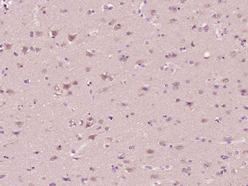
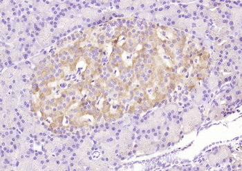
Paraformaldehyde-fixed, paraffin embedded (Human brain glioma), Antigen retrieval by boiling in sodium citrate buffer (pH6.0) for 15 min, Block endogenous peroxidase by 3% hydrogen peroxide for 20 minutes, Blocking buffer (normal goat serum) at 37°C for 30 min, Antibody incubation with (Bax) Polyclonal Antibody, Unconjugated (orb4655) at 1:400 overnight at 4°C, followed by operating according to SP Kit (Rabbit) instructionsand DAB staining.
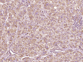
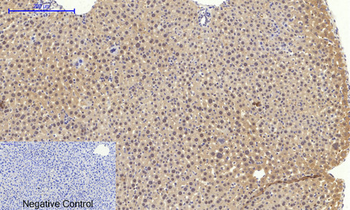
Paraformaldehyde-fixed, paraffin embedded (Human liver carcinoma), Antigen retrieval by boiling in sodium citrate buffer (pH6.0) for 15 min, Block endogenous peroxidase by 3% hydrogen peroxide for 20 minutes, Blocking buffer (normal goat serum) at 37°C for 30 min, Antibody incubation with (Bax) Polyclonal Antibody, Unconjugated (orb4655) at 1:400 overnight at 4°C, followed by operating according to SP Kit (Rabbit) instructionsand DAB staining.
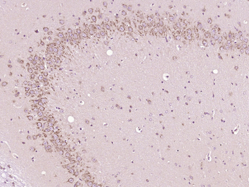
Paraformaldehyde-fixed, paraffin embedded (Rat brain), Antigen retrieval by boiling in sodium citrate buffer (pH6.0) for 15 min, Block endogenous peroxidase by 3% hydrogen peroxide for 20 minutes, Blocking buffer (normal goat serum) at 37°C for 30 min, Antibody incubation with (Bax) Polyclonal Antibody, Unconjugated (orb4655) at 1:400 overnight at 4°C, followed by operating according to SP Kit (Rabbit) instructionsand DAB staining.
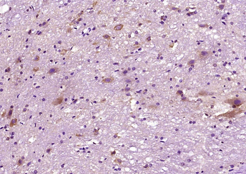
Paraformaldehyde-fixed, paraffin embedded (Rat spinal cord), Antigen retrieval by boiling in sodium citrate buffer (pH6.0) for 15 min, Block endogenous peroxidase by 3% hydrogen peroxide for 20 minutes, Blocking buffer (normal goat serum) at 37°C for 30 min, Antibody incubation with (Bax) Polyclonal Antibody, Unconjugated (orb4655) at 1:400 overnight at 4°C, followed by operating according to SP Kit (Rabbit) instructionsand DAB staining.
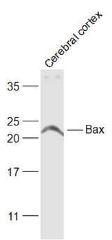
Sample: Cerebral cortex (Rat) Lysate at 40 ug, Primary: Anti-Bax (orb4655) at 1/1000 dilution, Secondary: IRDye800CW Goat Anti-Rabbit IgG at 1/20000 dilution, Predicted band size: 21 kD, Observed band size: 21 kD.
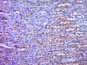
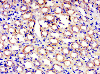
Tissue/Cell: rat stomach tissue, 4% Paraformaldehyde-fixed and paraffin-embedded, Antigen retrieval: citrate buffer (0.01M, pH 6.0), Boiling bathing for 15 min, Block endogenous peroxidase by 3% Hydrogen peroxide for 30 min, Blocking buffer (normal goat serum) at 37°C for 20 min, Incubation: Anti-Bax Polyclonal Antibody, Unconjugated (orb4655) 1:200, overnight at 4°C, followed by conjugation to the secondary antibody and DAB staining.
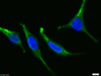
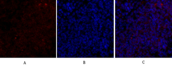
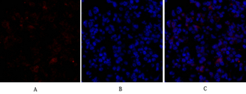
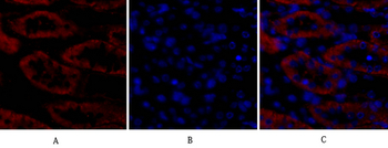
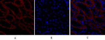
Tissue/Cell: Sh-sy5y cell, 4% Paraformaldehyde-fixed, Triton X-100 at room temperature for 20 min, Blocking buffer (normal goat serum) at 37°C for 20 min, Antibody incubation with (Bax) polyclonal Antibody, Unconjugated (orb4655) 1:100, 90 minutes at 37°C, followed by a FITC conjugated Goat Anti-Rabbit IgG antibody at 37°C for 90 minutes, DAPI (blue) was used to stain the cell nuclei.
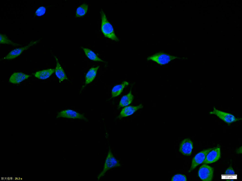
Tissue/Cell: Sh-sy5y cell, 4% Paraformaldehyde-fixed, Triton X-100 at room temperature for 20 min, Blocking buffer (normal goat serum) at 37°C for 20 min, Antibody incubation with (Bax) polyclonal Antibody, Unconjugated (orb4655) 1:100, 90 minutes at 37°C, followed by a FITC conjugated Goat Anti-Rabbit IgG antibody at 37°C for 90 minutes, DAPI (blue) was used to stain the cell nuclei.
Phospho-Bax (Ser184) Rabbit Polyclonal Antibody [orb4658]
ELISA, FC, IF, IHC-Fr, IHC-P, WB
Bovine, Canine, Porcine, Sheep
Human, Mouse, Rabbit, Rat
Rabbit
Polyclonal
Unconjugated
50 μl, 100 μl, 200 μlBax Polyclonal Antibody [orb1414573]
IF, IHC-P, WB
Human, Mouse, Rat
Rabbit
Polyclonal
Unconjugated
100 μlBax Antibody (BH3 Domain Specific) [orb1939544]
FC, WB
Human, Mouse
Rabbit
Polyclonal
Unconjugated
400 μlGRINA Rabbit Polyclonal Antibody [orb312256]
IF, IHC-Fr, IHC-P, WB
Bovine, Human, Rabbit, Sheep
Mouse, Rat
Rabbit
Polyclonal
Unconjugated
50 μl, 100 μl, 200 μl


