You have no items in your shopping cart.
Cart summary
Item 1 of 5
Item 1 of 5
Bax Antibody (BH3 Domain Specific)
Catalog Number: orb31066

| Catalog Number | orb31066 |
|---|---|
| Category | Antibodies |
| Description | Rabbit polyclonal antibody to BAX |
| Species/Host | Rabbit |
| Clonality | Polyclonal |
| Clone Number | RB0952 |
| Tested applications | FC, WB |
| Reactivity | Human, Mouse |
| Isotype | Rabbit IgG |
| Immunogen | Synthetic Peptide |
| Dilution range | WB: 1:2000, IHC-P: 1:50-100, FACS: 1:10-50 |
| Form/Appearance | Purified polyclonal antibody supplied in PBS with 0.09% (W/V) sodium azide. This antibody is purified through a protein A column, followed by peptide affinity purification. |
| Conjugation | Unconjugated |
| MW | 21184 |
| Target | BAX |
| UniProt ID | Q07812 |
| NCBI | NP_004315.1, NP_620119.2, NP_620118.1, NP_620116.1 |
| Storage | Maintain refrigerated at 2-8°C for up to 2 weeks. For long term storage store at -20°C in small aliquots to prevent freeze-thaw cycles |
| Alternative names | anti-Apoptosis regulator BAX antibody, anti- BAX a Read more... |
| Note | For research use only |
| Expiration Date | 12 months from date of receipt. |

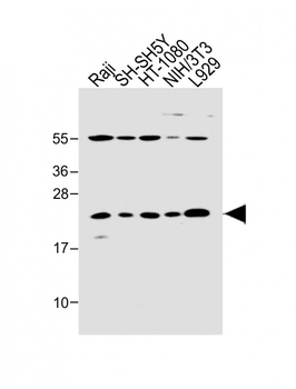
All lanes: Anti-Bax Antibody (BH3 Domain Specific) at 1:2000 dilution. Lane 1: HT-1080 whole cell lysate. Lane 2: Raji whole cell lysate. Lane 3: SH-SY5Y whole cell lysate. Lane 4: THP-1 whole cell lysate. Lane 5: NIH/3T3 whole cell lysate. Lane 6: L929 whole cell lysate.Lysates/proteins at 20 µg per lane. Secondary Goat Anti-Rabbit IgG, (H+L), Peroxidase conjugated at 1/10000 dilution. Predicted band size: 21 kDa. Blocking/Dilution buffer: 5% NFDM/TBST.

All lanes: Anti-Bax Antibody (BH3 Domain Specific) at 1:2000 dilution. Lane 1: Raji whole cell lysate. Lane 2: SH-SH5Y whole cell lysate. Lane 3: HT-1080 whole cell lysate. Lane 4: NIH/3T3 whole cell lysate. Lane 5: L929 whole cell lysate. Lysates/proteins at 20 µg per lane. Secondary Goat Anti-Rabbit IgG, (H+L), Peroxidase conjugated at 1/10000 dilution. Predicted band size: 21 kDa. Blocking/Dilution buffer: 5% NFDM/TBST.
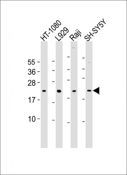
All lanes: Anti-Bax Antibody (BH3) at 1:2000 dilution. Lane 1: HT-1080 whole cell lysate. Lane 2: L929 whole cell lysate. Lane 3: Raji whole cell lysate. Lane 4: SH-SY5Y whole cell lysate.Lysates/proteins at 20 µg per lane. Secondary Goat Anti-Rabbit IgG, (H+L), Peroxidase conjugated at 1/10000 dilution. Predicted band size: 21 kDa. Blocking/Dilution buffer: 5% NFDM/TBST.
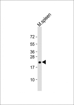
Anti-Bax Antibody (BH3) at 1:2000 dilution + mouse spleen lysate.Lysates/proteins at 20 µg per lane. Secondary Goat Anti-Rabbit IgG, (H+L), Peroxidase conjugated at 1/10000 dilution. Predicted band size: 21 kDa. Blocking/Dilution buffer: 5% NFDM/TBST.

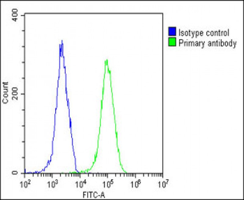
Overlay histogram showing HeLa cells (green line). The cells were fixed with 2% paraformaldehyde (10 min) and then permeabilized with 90% methanol for 10 min. The cells were then icubated in 2% bovine serum albumin to block non-specific protein-protein interactions followed by the antibody (1:25 dilution) for 60 min at 37°C. The secondary antibody used was Goat-Anti-Rabbit IgG, DyLight 488 Conjugated Highly Cross-Adsorbed at 1/200 dilution for 40 min at 37°C. Isotype control antibody (blue line) was rabbit IgG1 (1 μg/1x10^6 cells) used under the same conditions. Acquisition of > 10000 events was performed.
- Item 1 of 5
Bax Antibody (BH3 Domain Specific) [orb1939544]
FC, WB
Human, Mouse
Rabbit
Polyclonal
Unconjugated
400 μl 
Bax Antibody (BH3 Domain Specific) [orb1167959]
FC, IHC-P, WB
Human, Mouse
Rabbit
Polyclonal
Unconjugated
100 μl, 30 μl