You have no items in your shopping cart.
Cart summary
Item 1 of 10
Item 1 of 10
ARPC5 Recombinant Rabbit Monoclonal Antibody
Catalog Number: orb1499399

| Catalog Number | orb1499399 |
|---|---|
| Category | Antibodies |
| Description | ARPC5 Recombinant Rabbit Monoclonal Antibody |
| Species/Host | Rabbit |
| Clonality | Recombinant |
| Tested applications | ICC, IF, IHC-Fr, IHC-P, WB |
| Predicted Reactivity | Rat |
| Reactivity | Human, Mouse |
| Isotype | IgG |
| Immunogen | KLH conjugated synthetic peptide derived from human ARPC5 (1-50/151aa) |
| Antibody Type | Recombinant Antibody |
| Concentration | 1mg/ml |
| Dilution range | WB=1:500-2000, IHC-P=1:100-500, IHC-F=1:50, ICC/IF=1:50, IF=1:100-500 |
| Form/Appearance | Liquid |
| Conjugation | Unconjugated |
| MW | 16 kDa |
| Target | ARPC5 |
| UniProt ID | O15511 |
| Storage | Maintain refrigerated at 2-8°C for up to 2 weeks. For long term storage store at -20°C in small aliquots to prevent freeze-thaw cycles. |
| Buffer/Preservatives | 0.01M TBS (pH7.4) with 1% rAlbumin, 0.02% Proclin300 and 50% Glycerol. |
| Alternative names | ARPC5_HUMAN; Actin-related protein 2/3 complex sub Read more... |
| Note | For research use only |
| Expiration Date | 12 months from date of receipt. |

Blocking buffer: 5% NFDM/TBST, Primary Ab Dilution: 1:2000, Primary Ab incubation condition: 2 hours at room temperature, Secondary Ab: Goat Anti-Rabbit IgG H&L (HRP), Lysate: 1: HeLa, 2: HCT-116, 3: SW480, 4: Rat, brain, 5: Mouse brain, 6: Mouse ovary, Protein loading quantity: 20 µg, Exposure time: 60 s, Predicted MW: 16 kDa, Observed MW: 16 kDa.
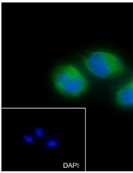
Cell line: Neuro-2a, Fixative: 4% Paraformaldehyde, Permeabilization: 0.1% TritonX-100, Primary Ab Dilution: 1:50, Primary incubation condition: 4°C overnight, Secondary Ab: Goat Anti-Rabbit IgG, Nuclear counter stain: DAPI (Blue), Comment: Color green is the positive signal for orb1499399.
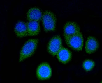
ICC staining of p16 ARC in N2A cells (green). Formalin fixed cells were permeabilized with 0.1% Triton X-100 in TBS for 10 minutes at room temperature and blocked with 10% negative goat serum for 15 minutes at room temperature. Cells were probed with the primary antibody (orb1499399, 1/50) for 1 hour at room temperature, washed with PBS. Alexa Fluor®488 conjugate-Goat anti-Rabbit IgG was used as the secondary antibody at 1/1000 dilution. The nuclear counter stain is DAPI (blue).
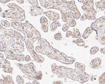
Immunohistochemical analysis of paraffin-embedded human placenta tissue using anti-p16 ARC antibody. The section was pre-treated using heat mediated antigen retrieval with Tris-EDTA buffer (pH9.0) for 20 minutes. The tissues were blocked in 1% BSA for 30 minutes at room temperature, washed with ddH2O and PBS, and then probed with the primary antibody (orb1499399, 1/50) for 30 minutes at room temperature. The detection was performed using an HRP conjugated compact polymer system. DAB was used as the chromogen. Tissues were counterstained with hematoxylin and mounted with DPX.
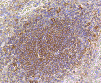
Immunohistochemical analysis of paraffin-embedded human spleen tissue using anti-p16 ARC antibody. The section was pre-treated using heat mediated antigen retrieval with Tris-EDTA buffer (pH9.0) for 20 minutes. The tissues were blocked in 1% BSA for 30 minutes at room temperature, washed with ddH2O and PBS, and then probed with the primary antibody (orb1499399, 1/50) for 30 minutes at room temperature. The detection was performed using an HRP conjugated compact polymer system. DAB was used as the chromogen. Tissues were counterstained with hematoxylin and mounted with DPX.
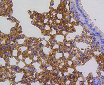
Immunohistochemical analysis of paraffin-embedded mouse lung tissue using anti-p16 ARC antibody. The section was pre-treated using heat mediated antigen retrieval with Tris-EDTA buffer (pH9.0) for 20 minutes. The tissues were blocked in 1% BSA for 30 minutes at room temperature, washed with ddH2O and PBS, and then probed with the primary antibody (orb1499399, 1/50) for 30 minutes at room temperature. The detection was performed using an HRP conjugated compact polymer system. DAB was used as the chromogen. Tissues were counterstained with hematoxylin and mounted with DPX.
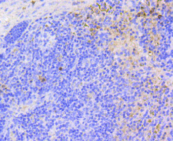
Immunohistochemical analysis of paraffin-embedded mouse spleen tissue using anti-p16 ARC antibody. The section was pre-treated using heat mediated antigen retrieval with Tris-EDTA buffer (pH9.0) for 20 minutes. The tissues were blocked in 1% BSA for 30 minutes at room temperature, washed with ddH2O and PBS, and then probed with the primary antibody (orb1499399, 1/50) for 30 minutes at room temperature. The detection was performed using an HRP conjugated compact polymer system. DAB was used as the chromogen. Tissues were counterstained with hematoxylin and mounted with DPX.
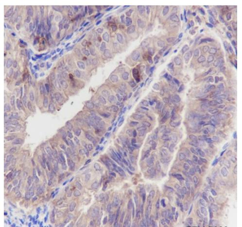
Tissue: Human endometrium carcinoma, Section type: Formalin fixed & Paraffin embedded section, Retrieval method: High temperature and high pressure, Retrieval buffer: Tris/EDTA buffer, pH9.0, Primary Ab Dilution: 1:100, Primary Ab incubation condition: 1 hour at room temperature, Secondary Ab: Anti-Rabbit and Mouse, Polymer HRP (Ready to use), Counter stain: Hematoxylin (Blue), Comment: Color brown is the positive signal for orb1499399.
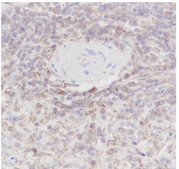
Tissue: Human spleen, Section type: Formalin fixed & Paraffin embedded section, Retrieval method: High temperature and high pressure, Retrieval buffer: Tris/EDTA buffer, pH9.0, Primary Ab Dilution: 1:100, Primary Ab incubation condition: 1 hour at room temperature, Secondary Ab: Anti-Rabbit and Mouse, Polymer HRP (Ready to use), Counter stain: Hematoxylin (Blue), Comment: Color brown is the positive signal for orb1499399.
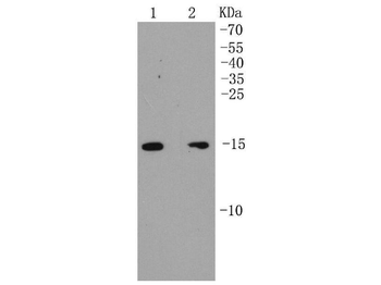
Western blot analysis of p16 ARC on different lysates. Proteins were transferred to a PVDF membrane and blocked with 5% BSA in PBS for 1 hour at room temperature. The primary antibody (orb1499399, 1/500) was used in 5% BSA at room temperature for 2 hours. Goat Anti-Rabbit IgG - HRP Secondary Antibody (HA1001) at 1:200000 dilution was used for 1 hour at room temperature. Positive control: Lane 1: MCF-7 cell lysate, Lane 2: SK-Br-3 cell lysate.
