You have no items in your shopping cart.
Cart summary
Item 1 of 2
Item 1 of 2
ARFGAP3 antibody
Catalog Number: orb750535

| Catalog Number | orb750535 |
|---|---|
| Category | Antibodies |
| Description | ARFGAP3 antibody |
| Species/Host | Rabbit |
| Clonality | Polyclonal |
| Tested applications | ELISA, IF, IP, WB |
| Reactivity | Human |
| Isotype | Antiserum |
| Immunogen | This whole rabbit serum was prepared by repeated immunizations with a truncated recombinant sequence of ArfGAP3 fused to GST. |
| Concentration | 70mg/mL |
| Dilution range | ELISA: 1:5,000, IF: 1:100, IP: User Optimized, WB: 1:1,000 |
| Form/Appearance | Liquid (sterile filtered) |
| Purity | Anti-ArfGAP3 antibody was prepared from monospecific antiserum by delipidation and defibrination. Further purification was used to remove the GST tag. The antibody detects ArfGAP3 in cell lysates. A BLAST analysis was used to suggest cross reactivity with human, monkey, and orangutan for ArfGAP3. Cross-reactivity with AfGAP3 from other sources have not been determined. |
| Conjugation | Unconjugated |
| UniProt ID | Q9NP61 |
| NCBI | NP_001135765.1 |
| Storage | Store vial at -20° C or below prior to opening. This vial contains a relatively low volume of reagent (25 µL). To minimize loss of volume dilute 1:10 by adding 225 µL of the buffer stated above directly to the vial. Recap, mix thoroughly and briefly centrifuge to collect the volume at the bottom of the vial. Use this intermediate dilution when calculating final dilutions as recommended below. Store the vial at -20°C or below after dilution. Avoid cycles of freezing and thawing. |
| Buffer/Preservatives | 0.01% (w/v) Sodium Azide. 0.02 M Potassium Phosphate, 0.15 M Sodium Chloride, pH 7.2 |
| Alternative names | rabbit anti-ArfGAP3 Antibody, ARFGAP1, ADP-ribosyl Read more... |
| Note | For research use only |
| Application notes | ArfGAP3 has been tested for use in Immunofluorescence and western blotting. Specific conditions for reactivity should be optimized by the end user. Expect a band approximately 57 kDa in size by western blotting in the appropriate cell lysate or extract. |
| Expiration Date | 12 months from date of receipt. |
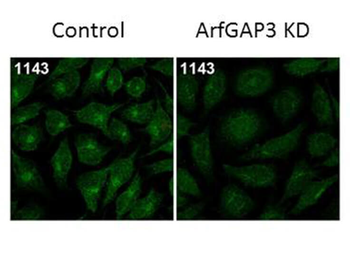
Immunofluorescence Microscopy of Rabbit Anti-ArfGAP3 Antibody. Tissue: HeLa Whole Cell. Fixation: MeOH. Antigen retrieval: not required. Primary antibody: ArfGAP3 antibody at 1:100 for 1 h at RT. Secondary antibody: Fluorescein rabbit secondary antibody at 1:10000 for 45 min at RT. Localization: ArfGAP3 is cytoplasmic. Staining: ArfGAP3 as green fluorescent signal.

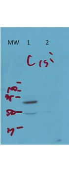
Western Blot of Rabbit Anti-ArfGAP3 Antibody. Lane 1 (C): HeLa Whole Cell. Lane 2 (si): HeLa Whole Cell siRNA treated. Load: 10 µg per lane. Primary antibody: ArfGAP3 antibody at 1:1000 for overnight at 4°C. Secondary antibody: IRDye800™ rabbit secondary antibody at 1:10000 for 45 min at RT. Block: 5% BLOTTO overnight at 4°C. Predicted/Observed size: 57 kDa for endogenous Arf-GAP3. Other band(s): non-specific band ~50 kDa.
- Item 1 of 4
- Item 1 of 4
- Item 1 of 2
- Item 1 of 2
- Item 1 of 1


Submit a review
Filter by Rating

- 5 stars

- 4 stars
- 3 stars
- 2 stars
- 1 stars
