You have no items in your shopping cart.
Cart summary
Item 1 of 2
Item 1 of 2
ARFGAP3 antibody
Catalog Number: orb750534

| Catalog Number | orb750534 |
|---|---|
| Category | Antibodies |
| Description | ARFGAP3 antibody |
| Species/Host | Rabbit |
| Clonality | Polyclonal |
| Tested applications | ELISA, IF, IP, WB |
| Reactivity | Human |
| Isotype | Antiserum |
| Immunogen | This whole rabbit serum was prepared by repeated immunizations with a truncated recombinant sequence of ArfGAP3 fused to GST. |
| Concentration | 70mg/mL |
| Dilution range | ELISA: 1:5,000, IF: 1:100, IP: User Optimized, WB: 1:1,000 |
| Form/Appearance | Liquid (sterile filtered) |
| Purity | Anti-ArfGAP3 antibody was prepared from monospecific antiserum by delipidation and defibrination. Further purification was used to remove the GST tag. The antibody detects ArfGAP3 in cell lysates. A BLAST analysis was used to suggest cross reactivity with human, monkey, and orangutan for ArfGAP3. Cross-reactivity with AfGAP3 from other sources have not been determined. |
| Conjugation | Unconjugated |
| UniProt ID | Q9NP61 |
| NCBI | NP_001135765.1 |
| Storage | Store vial at -20° C prior to opening. Aliquot contents and freeze at -20° C or below for extended storage. Avoid cycles of freezing and thawing. Centrifuge product if not completely clear after standing at room temperature. This product is stable for several weeks at 4° C as an undiluted liquid. Dilute only prior to immediate use. |
| Buffer/Preservatives | 0.01% (w/v) Sodium Azide. 0.02 M Potassium Phosphate, 0.15 M Sodium Chloride, pH 7.2 |
| Alternative names | rabbit anti-ArfGAP3 Antibody, ARFGAP1, ADP-ribosyl Read more... |
| Note | For research use only |
| Application notes | ArfGAP3 has been tested for use in Immunofluorescence and western blotting. Specific conditions for reactivity should be optimized by the end user. Expect a band approximately 57 kDa in size by western blotting in the appropriate cell lysate or extract. |
| Expiration Date | 12 months from date of receipt. |

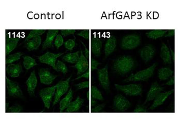
Immunofluorescence Microscopy of Rabbit Anti-ArfGAP3 Antibody. Tissue: HeLa Whole Cell. Fixation: MeOH. Antigen retrieval: not required. Primary antibody: ArfGAP3 antibody at 1:100 for 1 h at RT. Secondary antibody: Fluorescein rabbit secondary antibody at 1:10000 for 45 min at RT. Localization: ArfGAP3 is cytoplasmic. Staining: ArfGAP3 as green fluorescent signal.
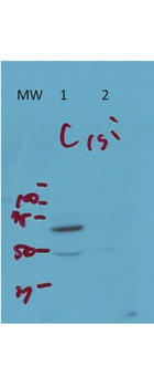
Western Blot of Rabbit Anti-ArfGAP3 Antibody. Lane 1 (C): HeLa Whole Cell. Lane 2 (si): HeLa Whole Cell siRNA treated. Load: 10 µg per lane. Primary antibody: ArfGAP3 antibody at 1:1000 for overnight at 4°C. Secondary antibody: IRDye800™ rabbit secondary antibody at 1:10000 for 45 min at RT. Block: 5% BLOTTO overnight at 4°C. Predicted/Observed size: 57 kDa for endogenous Arf-GAP3. Other band(s): non-specific band ~50 kDa.
- Item 1 of 4
- Item 1 of 4
- Item 1 of 2
- Item 1 of 2
- Item 1 of 1


Submit a review
Filter by Rating

- 5 stars

- 4 stars
- 3 stars
- 2 stars
- 1 stars
