You have no items in your shopping cart.
Cart summary
Item 1 of 5
Item 1 of 5
APP Antibody
Catalog Number: orb1239195

| Catalog Number | orb1239195 |
|---|---|
| Category | Antibodies |
| Description | APP Antibody |
| Species/Host | Rabbit |
| Clonality | Polyclonal |
| Tested applications | ELISA, IF, IHC-P, WB |
| Predicted Reactivity | Guinea pig, Porcine |
| Reactivity | Human, Mouse, Rat |
| Isotype | IgG |
| Immunogen | APP antibody was raised against a 15 amino acid peptide near the carboxy terminus of human APP.The immunogen is located within the last 50 amino acids of APP. |
| Concentration | 1 mg/ml |
| Dilution range | APP antibody can be used for detection of APP and the C99 fragment by Western blot at 0.5 - 1 μg/mL. Antibody can also be used for immunohistochemistry starting at 2 μg/mL. For immunofluorescence start at 20 μg/mL.Antibody validated: Western Blot in mouse samples; Immunohistochemistry in mouse and rat samples and Immunofluorescence in mouse and rat samples. All other applications and species not yet tested. |
| Form/Appearance | Liquid |
| Conjugation | Unconjugated |
| MW | Predicted: 83 kDa Observed: 110 kDa |
| Target | APP |
| UniProt ID | P05067 |
| NCBI | CAA30050 |
| Storage | APP antibody can be stored at 4°C up to one year. Antibodies should not be exposed to prolonged high temperatures. |
| Buffer/Preservatives | APP Antibody is supplied in PBS containing 0.02% sodium azide. |
| Alternative names | APP Antibody: AAA, AD1, PN2, ABPP, APPI, CVAP, ABE Read more... |
| Note | For research use only |
| Application notes | APP antibody can be used for detection of APP and the C99 fragment by Western blot at 0.5 - 1 μg/mL. Antibody can also be used for immunohistochemistry starting at 2 μg/mL. For immunofluorescence start at 20 μg/mL.Antibody validated: Western Blot in mouse samples; Immunohistochemistry in mouse and rat samples and Immunofluorescence in mouse and rat samples. All other applications and species not yet tested. |
| Expiration Date | 12 months from date of receipt. |

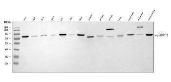
Western blot analysis of APP in mouse brain tissue lysate with APP antibody at (A) 0.5 and (B) 1 µg/ml.

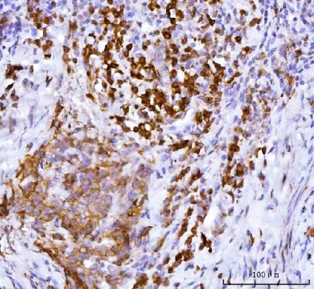
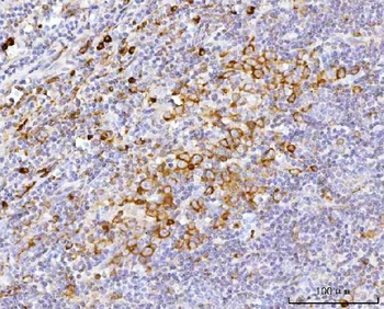
Immunohistochemical staining of rat brain using APP antibody at 2 µg/mL.

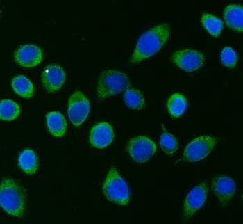
Immunofluorescence of APP in Rat Brain cells with APP antibody at 20 µg/mL.

Immunohistochemistry of APP in mouse brain tissue with APP Antibody at 5 µg/mL.

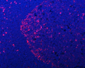
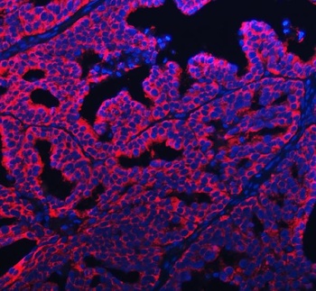
Immunofluorescence of APP in mouse brain tissue with APP Antibody at 20 µg/mL. Green: APP antibody (orb1239195) Red: Phylloidin staining Blue: DAPI staining.
- Item 1 of 8
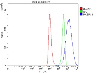
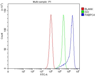
APP-1/PABPC4 Antibody [orb1173479]
FC, ICC, IF, IHC, WB
Human, Mouse, Rat
Rabbit
Polyclonal
Unconjugated
10 μg, 100 μg - Item 1 of 7
beta Amyloid/APP Antibody [orb196261]
FC, ICC, IF, IHC, IHC-Fr, WB
Bovine, Equine, Monkey, Rabbit
Human, Mouse, Rat
Rabbit
Polyclonal
Unconjugated
10 μg, 100 μg - Item 1 of 6
Cathepsin B antibody [orb240235]
ELISA, IF, IHC, WB
Human, Mouse
Rabbit
Polyclonal
Unconjugated
100 μg, 50 μg - Item 1 of 6
Amyloid beta A4 protein antibody [orb242610]
ELISA, IF, IHC, WB
Human, Mouse
Rabbit
Polyclonal
Unconjugated
50 μg, 100 μg - Item 1 of 6


Submit a review
Filter by Rating

- 5 stars

- 4 stars
- 3 stars
- 2 stars
- 1 stars
