You have no items in your shopping cart.
Cart summary
Item 1 of 8
Item 1 of 8
APE1/APEX1 Antibody
Catalog Number: orb215883

| Catalog Number | orb215883 |
|---|---|
| Category | Antibodies |
| Description | APE1/APEX1 Antibody |
| Species/Host | Rabbit |
| Clonality | Polyclonal |
| Tested applications | FC, ICC, IF, IHC, WB |
| Reactivity | Human, Mouse, Rat |
| Isotype | Rabbit IgG |
| Immunogen | E.coli-derived human APE1 recombinant protein (Position: P2-L318). Human APE1 shares 94% and 93% amino acid (aa) sequences identity with mouse and rat APE1, respectively. |
| Concentration | Adding 0.2 ml of distilled water will yield a concentration of 500 μg/ml. |
| Form/Appearance | Lyophilized |
| Conjugation | Unconjugated |
| MW | 35555 MW |
| UniProt ID | P27695 |
| Storage | Store at -20˚C for one year from date of receipt. After reconstitution, at 4˚C for one month. It can also be aliquotted and stored frozen at -20˚C for six months. Avoid repeated freeze-thaw cycles. |
| Alternative names | DNA- (apurinic or apyrimidinic site) lyase;3.1.-.- Read more... |
| Note | For research use only |
| Application notes | WB: The detection limit for APE1 is approximately 0.25ng/lane under reducing conditions. Tested Species: In-house tested species with positive results. By Heat: Boiling the paraffin sections in 10mM citrate buffer, pH6.0, for 20mins is required for the staining of formalin/paraffin sections. Other applications have not been tested. Optimal dilutions should be determined by end users. . Add 0.2ml of distilled water will yield a concentration of 500ug/ml. |
| Expiration Date | 12 months from date of receipt. |

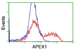
Flow Cytometry analysis of U937 cells using anti-APEX1 antibody (Blue line).Isotype control antibody (Green line) was rabbit IgG .Unlabelled sample (Red line) was also used as a control.

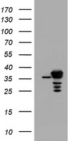
WB analysis of APEX1 using anti-APEX1 antibody.Lane 1:Recombinant Human APEX1 Protein 0.5ng.

WB analysis of APEX1 using anti-APEX1 antibody.Lane 1:NRK Cell;2:HELA Cell;3:PC-12 Cell;4:RH35 Cell;5:HEPA Cell;6:MCF Cell;7:A549 Cell;8:Human Placenta Tissue;9:A431 Cell.

IF analysis of APEX1 using anti-APEX1 antibody.APEX1 was detected in immunocytochemical section of U20S cell.

IF analysis of APEX1 using anti-APEX1 antibody. APEX1 was detected in paraffin-embedded section of human colon cancer tissue.

IHC analysis of APEX1 using anti-APEX1 antibody.APEX1 was detected in paraffin-embedded section of human lung cancer tissue.

IHC analysis of APEX1 using anti-APEX1 antibody.APEX1 was detected in paraffin-embedded section of mouse brain tissue.

IHC analysis of APEX1 using anti-APEX1 antibody.APEX1 was detected in paraffin-embedded section of rat brain tissue.
- Item 1 of 3
APE1/APEX1 Antibody [orb47961]
IHC, WB
Hamster
Human, Mouse, Rat
Rabbit
Polyclonal
Unconjugated
10 μg, 100 μg - Item 1 of 2
APEX1 Antibody [orb1248020]
ELISA, IHC, WB
Bovine, Canine, Porcine
Human
Goat
Polyclonal
Unconjugated
0.1 mg - Item 1 of 2
APE1 APEX1 Antibody (monoclonal, 5C11) [orb421116]
FC, ICC, IHC, WB
Human
Mouse
Monoclonal
Unconjugated
10 μg, 100 μg - Item 1 of 1
APE1 (APEX1) antibody [orb1332988]
IHC, WB
Canine, Human, Monkey, Mouse, Rat
Mouse
Monoclonal
Unconjugated
100 μg - Item 1 of 1
APE1 (APEX1) antibody [orb1332989]
FC, IF, IHC, WB
Human, Mouse, Rat
Mouse
Monoclonal
Unconjugated
100 μg


Submit a review
Filter by Rating

- 5 stars

- 4 stars
- 3 stars
- 2 stars
- 1 stars
