You have no items in your shopping cart.
Cart summary
Item 1 of 6
Item 1 of 6
Anti-YB1/YBX1 Antibody
Catalog Number: orb76271

| Catalog Number | orb76271 |
|---|---|
| Category | Antibodies |
| Description | Anti-YB1/YBX1 Antibody. Tested in Flow Cytometry, IHC, WB applications. This antibody reacts with Human, Mouse, Rat. |
| Species/Host | Rabbit |
| Clonality | Polyclonal |
| Tested applications | FC, IHC, WB |
| Reactivity | Human, Mouse, Rat |
| Isotype | Rabbit IgG |
| Immunogen | A synthetic peptide corresponding to a sequence in the middle region of human YB1, identical to the related rat and mouse sequences. |
| Antibody Type | Primary Antibody |
| Concentration | Adding 0.2 ml of distilled water will yield a concentration of 500 μg/ml. |
| Form/Appearance | Lyophilized |
| Conjugation | Unconjugated |
| MW | 50 kDa |
| UniProt ID | P67809 |
| Storage | Maintain refrigerated at 2-8°C for up to 2 weeks. For long term storage store at -20°C in small aliquots to prevent freeze-thaw cycles. |
| Alternative names | Nuclease-sensitive element-binding protein 1; CCAA Read more... |
| Note | For research use only |
| Application notes | Western blot, 0.1-0.5μg/ml, Human, Mouse, Rat Immunohistochemistry(Paraffin-embedded Section), 2-5 μg/ml, Human, Mouse, Rat Flow Cytometry (Fixed), 1-3 μg/1x106 cells, Human. Add 0.2ml of distilled water will yield a concentration of 500ug/ml |
| Expiration Date | 12 months from date of receipt. |

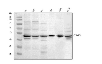
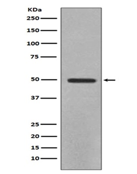
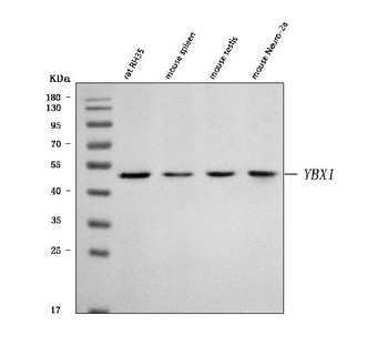
Western blot analysis of YBX1 using anti-YBX1 antibody (orb76271). Electrophoresis was performed on a 5-20% SDS-PAGE gel at 70V (Stacking gel)/90V (Resolving gel) for 2-3 hours. The sample well of each lane was loaded with 30 ug of sample under reducing conditions. Lane 1: human MCF-7 whole cell lysates, Lane 2: human 293T whole cell lysates, Lane 3: human HeLa whole cell lysates, Lane 4: human Jurkat whole cell lysates, Lane 5: human T47D whole cell lysates, Lane 6: human THP-1 whole cell lysates, Lane 7: human MOLT4 whole cell lysates, Lane 8: human HL-60 whole cell lysates, Lane 9: rat testis tissue lysates, Lane 10: mouse stomach tissue lysates, Lane 11: mouse testis tissue lysates. After electrophoresis, proteins were transferred to a nitrocellulose membrane at 150 mA for 50-90 minutes. Blocked the membrane with 5% non-fat milk/TBS for 1.5 hour at RT. The membrane was incubated with rabbit anti-YBX1 antigen affinity purified polyclonal antibody (Catalog # orb76271) at 0.5 μg/mL overnight at 4°C, then washed with TBS-0.1%Tween 3 times with 5 minutes each and probed with a goat anti-rabbit IgG-HRP secondary antibody at a dilution of 1:5000 for 1.5 hour at RT. The signal is developed using an Enhanced Chemiluminescent detection (ECL) kit (Catalog # orb90503) with Tanon 5200 system. A specific band was detected for YBX1 at approximately 50 kDa. The expected band size for YBX1 is at 36 kDa.

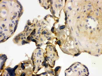
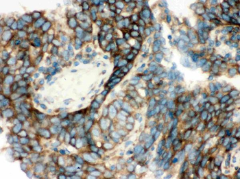
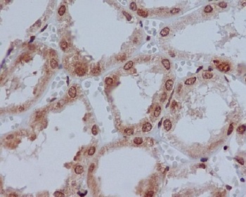
IHC analysis of YBX1 using anti-YBX1 antibody (orb76271). YBX1 was detected in a paraffin-embedded section of human rectal cancer tissue. Heat mediated antigen retrieval was performed in EDTA buffer (pH 8.0, epitope retrieval solution). The tissue section was blocked with 10% goat serum. The tissue section was then incubated with 2 μg/ml rabbit anti-YBX1 Antibody (orb76271) overnight at 4°C. Peroxidase Conjugated Goat Anti-rabbit IgG was used as secondary antibody and incubated for 30 minutes at 37°C. The tissue section was developed using HRP Conjugated Rabbit IgG Super Vision Assay Kit with DAB as the chromogen.

IHC analysis of YBX1 using anti-YBX1 antibody (orb76271). YBX1 was detected in a paraffin-embedded section of human ovarian cancer tissue. Heat mediated antigen retrieval was performed in EDTA buffer (pH 8.0, epitope retrieval solution). The tissue section was blocked with 10% goat serum. The tissue section was then incubated with 2 μg/ml rabbit anti-YBX1 Antibody (orb76271) overnight at 4°C. Peroxidase Conjugated Goat Anti-rabbit IgG was used as secondary antibody and incubated for 30 minutes at 37°C. The tissue section was developed using HRP Conjugated Rabbit IgG Super Vision Assay Kit with DAB as the chromogen.

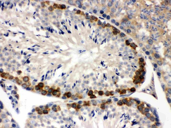
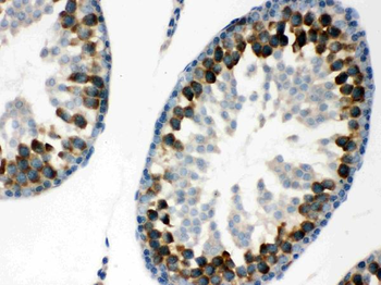
IHC analysis of YBX1 using anti-YBX1 antibody (orb76271). YBX1 was detected in a paraffin-embedded section of mouse testis tissue. Heat mediated antigen retrieval was performed in EDTA buffer (pH 8.0, epitope retrieval solution). The tissue section was blocked with 10% goat serum. The tissue section was then incubated with 2 μg/ml rabbit anti-YBX1 Antibody (orb76271) overnight at 4°C. Peroxidase Conjugated Goat Anti-rabbit IgG was used as secondary antibody and incubated for 30 minutes at 37°C. The tissue section was developed using HRP Conjugated Rabbit IgG Super Vision Assay Kit with DAB as the chromogen.

IHC analysis of YBX1 using anti-YBX1 antibody (orb76271). YBX1 was detected in a paraffin-embedded section of rat testis tissue. Heat mediated antigen retrieval was performed in EDTA buffer (pH 8.0, epitope retrieval solution). The tissue section was blocked with 10% goat serum. The tissue section was then incubated with 2 μg/ml rabbit anti-YBX1 Antibody (orb76271) overnight at 4°C. Peroxidase Conjugated Goat Anti-rabbit IgG was used as secondary antibody and incubated for 30 minutes at 37°C. The tissue section was developed using HRP Conjugated Rabbit IgG Super Vision Assay Kit with DAB as the chromogen.

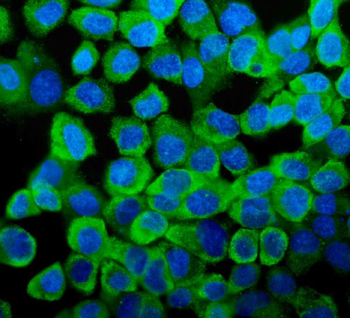
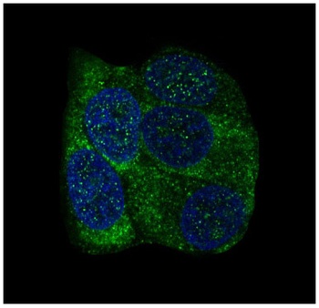
Flow Cytometry analysis of HEL cells using anti-YBX1 antibody (orb76271). Overlay histogram showing HEL cells stained with orb76271 (Blue line). The cells were blocked with 10% normal goat serum. And then incubated with rabbit anti-YBX1 Antibody (orb76271, 1 μg/1x10^6 cells) for 30 min at 20°C. DyLight®488 conjugated goat anti-rabbit IgG (5-10 μg/1x10^6 cells) was used as secondary antibody for 30 minutes at 20°C. Isotype control antibody (Green line) was rabbit IgG (1 μg/1x10^6) used under the same conditions. Unlabelled sample (Red line) was also used as a control.
- Item 1 of 7
Anti-YB1/YBX1 Antibody [orb259642]
FC, ICC, IF, IHC, IHC-Fr, WB
Human, Mouse, Rat
Rabbit
Polyclonal
Unconjugated
10 μg, 100 μg - Item 1 of 3
Anti-YB1 YBX1 Rabbit Monoclonal Antibody [orb547590]
FC, ICC, IF, IHC, IP, WB
Human, Mouse, Rat
Rabbit
Monoclonal
Unconjugated
30 μl, 100 μl - Item 1 of 2

Anti-YB1/YBX1 Antibody [orb2621442]
FC, ICC, IF, IHC, IHC-Fr, WB
Human, Mouse, Rat
Rabbit
Polyclonal
iFluor647
100 μgAnti-YB1/YBX1 Antibody [orb2621443]
FC, ICC, IF, IHC, IHC-Fr, WB
Human, Mouse, Rat
Rabbit
Polyclonal
PE
100 μg