You have no items in your shopping cart.
Cart summary
Item 1 of 6
Item 1 of 6
Anti-Von Willebrand Factor/VWF Antibody
Catalog Number: orb234319

| Catalog Number | orb234319 |
|---|---|
| Category | Antibodies |
| Description | Anti-Von Willebrand Factor/VWF Antibody |
| Species/Host | Rabbit |
| Clonality | Polyclonal |
| Tested applications | IF, IHC, WB |
| Reactivity | Mouse, Rat |
| Isotype | Rabbit IgG |
| Immunogen | E.coli-derived mouse VWF recombinant protein (Position: M1304-E1452). |
| Concentration | Adding 0.2 ml of distilled water will yield a concentration of 500 μg/ml. |
| Form/Appearance | Lyophilized |
| Conjugation | Unconjugated |
| MW | 309 kDa |
| UniProt ID | Q8CIZ8 |
| Storage | Maintain refrigerated at 2-8°C for up to 2 weeks. For long term storage store at -20°C in small aliquots to prevent freeze-thaw cycles. |
| Alternative names | von Willebrand factor; vWF; von Willebrand antigen Read more... |
| Note | For research use only |
| Application notes | Western blot, 0.1-0.5μg/ml, Mouse, Rat Immunohistochemistry (Paraffin-embedded Section), 0.5-1μg/ml, Mouse, Rat Immunofluorescence, 2μg/ml, Rat. Add 0.2ml of distilled water will yield a concentration of 500ug/ml |
| Expiration Date | 12 months from date of receipt. |

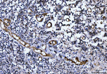
Anti-VWF Picoband antibody, IHC(P): Mouse Liver Tissue.
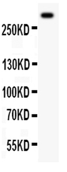
Anti-VWF Picoband antibody, Western blotting. All lanes: Anti VWF at 0.5 ug/ml. WB: Mouse Lung Tissue Lysate at 50 ug. Predicted bind size: 309 KD. Observed bind size: 309 KD.
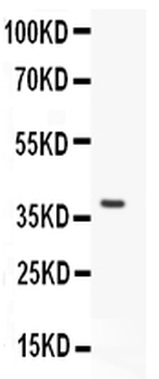
Anti-VWF Picoband antibody, Western blotting. All lanes: Anti VWF at 0.5 ug/ml. WB: Recombinant Mouse VWF Protein 0.5 ng. Predicted bind size: 37 KD. Observed bind size: 37 KD.
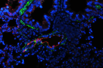
IF analysis of VWF and alpha-Smooth Muscle Actin using anti-VWF antibody and anti-alpha-Smooth Muscle Actin antibody. VWF and alpha-Smooth Muscle Actin a paraffin-embedded section of rat lung tissue. Heat mediated antigen retrieval was performed in EDTA buffer (pH8.0, epitope retrieval solution). The tissue section was blocked with 10% goat serum. The tissue section was then incubated with 2 µg/mL rabbit anti-VWF antibody and mouse anti-alpha-Smooth Muscle Actin Antibody overnight at 4°C. DyLight®488 Conjugated Goat Anti-Rabbit IgG and Cy3 Conjugated Goat Anti-Mouse IgG were used as secondary antibody at 1:100 dilution and incubated for 30 minutes at 37°C. The section was counterstained with DAPI. Visualize using a fluorescence microscope and filter sets appropriate for the label used.
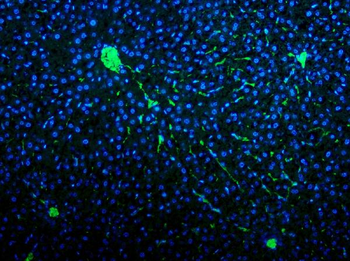
IF analysis of VWF using anti-VWF antibody. VWF was detected in paraffin-embedded section of rat liver tissue. Heat mediated antigen retrieval was performed in EDTA buffer (pH8.0, epitope retrieval solution). The tissue section was blocked with 10% goat serum. The tissue section was then incubated with 2 µg/mL rabbit anti-VWF Antibody overnight at 4°C. DyLight®488 Conjugated Goat Anti-Rabbit IgG was used as secondary antibody at 1:100 dilution and incubated for 30 minutes at 37°C. The section was counterstained with DAPI. Visualize using a fluorescence microscope and filter sets appropriate for the label used.
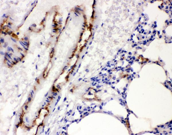
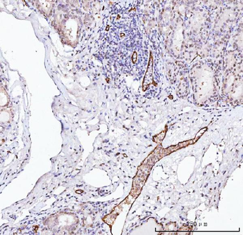
IHC analysis of VWF using anti-VWF antibody. VWF was detected in paraffin-embedded section of rat lung tissues. Heat mediated antigen retrieval was performed in citrate buffer (pH6, epitope retrieval solution) for 20 mins. The tissue section was blocked with 10% goat serum. The tissue section was then incubated with 1 µg/ml rabbit anti-VWF Antibody overnight at 4°C. Biotinylated goat anti-rabbit IgG was used as secondary antibody and incubated for 30 minutes at 37°C. The tissue section was developed using Strepavidin-Biotin-Complex (SABC) with DAB as the chromogen.
- Item 1 of 4
Anti-Von Willebrand Factor/VWF Antibody [orb182394]
FC, ICC, IHC, IHC-Fr, WB
Human
Rabbit
Polyclonal
Unconjugated
10 μg, 100 μg - Item 1 of 2
Anti-Von Willebrand Factor Antibody [orb1289887]
IH, WB
Human, Mouse, Rat
Rabbit
Polyclonal
Unconjugated
30 μl, 100 μl, 200 μl - Item 1 of 2
Anti-Von Willebrand Factor Antibody [orb1474085]
FC, WB
Human
Mouse
Monoclonal
Unconjugated
200 μl, 100 μl, 50 μl - Item 1 of 3
Anti-Von Willebrand Factor/VWF Antibody [orb1152307]
ELISA, IHC
Human
Rabbit
Polyclonal
Unconjugated
10 μg, 100 μg - Item 1 of 1
Anti-Von Willebrand Factor Antibody [orb1421650]
WB
Human, Mouse, Rat
Rabbit
Polyclonal
Unconjugated
100 μl