You have no items in your shopping cart.
Cart summary
Item 1 of 26
Item 1 of 26
Anti-Musashi 1/Msi1 Antibody Picoband
Catalog Number: orb19089

| Catalog Number | orb19089 |
|---|---|
| Category | Antibodies |
| Description | Anti-Musashi 1/Msi1 Antibody. Tested in IF, IHC, ICC, WB applications. This antibody reacts with Human, Mouse, Rat. |
| Species/Host | Rabbit |
| Clonality | Polyclonal |
| Tested applications | FC, ICC, IF, IHC, WB |
| Reactivity | Human, Mouse, Rat |
| Isotype | Rabbit IgG |
| Immunogen | A synthetic peptide corresponding to a sequence at the N-terminus of human Musashi 1/Msi1, identical to the related mouse and rat sequences. |
| Concentration | Adding 0.2 ml of distilled water will yield a concentration of 500 μg/ml. |
| Form/Appearance | Lyophilized |
| Conjugation | Unconjugated |
| MW | 39 kDa |
| UniProt ID | O43347 |
| Storage | Maintain refrigerated at 2-8°C for up to 2 weeks. For long term storage store at -20°C in small aliquots to prevent freeze-thaw cycles. |
| Alternative names | RNA-binding protein Musashi homolog 1; Musashi-1; Read more... |
| Note | For research use only |
| Application notes | Western blot, 0.1-0.5μg/ml, Human, Mouse, Rat Immunohistochemistry (Paraffin-embedded Section), 0.5-1μg/ml, Human, Mouse, Rat, By Heat Immunocytochemistry/Immunofluorescence, 5 μg/ml, Human Flow Cytometry (Fixed), 1-3 μg/1x106 cells, Human. Add 0.2ml of distilled water will yield a concentration of 500ug/ml |
| Expiration Date | 12 months from date of receipt. |

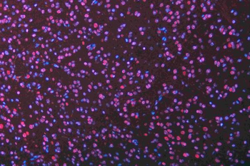
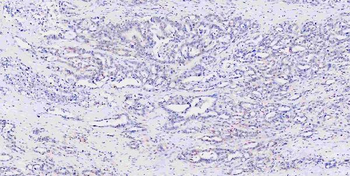
IF analysis of Musashi 1/Msi1 using anti-Musashi 1/Msi1 antibody. Musashi 1/Msi1 was detected in a paraffin-embedded section of human breast cancer tissue. Heat mediated antigen retrieval was performed in EDTA buffer (pH8.0, epitope retrieval solution). The tissue section was blocked with 10% goat serum. The tissue section was then incubated with 25 µg/mL rabbit anti-Musashi 1/Msi1 Antibody overnight at 4°C. DyLight®594 Conjugated Goat Anti-Rabbit IgG was used as secondary antibody at 1:100 dilution and incubated for 30 minutes at 37°C. The section was counterstained with DAPI. Visualize using a fluorescence microscope and filter sets appropriate for the label used.
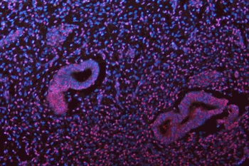
IF analysis of Musashi 1/Msi1 using anti-Musashi 1/Msi1 antibody. Musashi 1/Msi1 was detected in a paraffin-embedded section of human endometrial cancer tissue. Heat mediated antigen retrieval was performed in EDTA buffer (pH8.0, epitope retrieval solution). The tissue section was blocked with 10% goat serum. The tissue section was then incubated with 25 µg/mL rabbit anti-Musashi 1/Msi1 Antibody overnight at 4°C. DyLight®594 Conjugated Goat Anti-Rabbit IgG was used as secondary antibody at 1:100 dilution and incubated for 30 minutes at 37°C. The section was counterstained with DAPI. Visualize using a fluorescence microscope and filter sets appropriate for the label used.
IF analysis of Musashi 1/Msi1 using anti-Musashi 1/Msi1 antibody. Musashi 1/Msi1 was detected in a paraffin-embedded section of human pancreatic cancer tissue. Heat mediated antigen retrieval was performed in EDTA buffer (pH8.0, epitope retrieval solution). The tissue section was blocked with 10% goat serum. The tissue section was then incubated with 25 µg/mL rabbit anti-Musashi 1/Msi1 Antibody overnight at 4°C. DyLight®594 Conjugated Goat Anti-Rabbit IgG was used as secondary antibody at 1:100 dilution and incubated for 30 minutes at 37°C. The section was counterstained with DAPI. Visualize using a fluorescence microscope and filter sets appropriate for the label used.
IF analysis of Musashi 1/Msi1 using anti-Musashi 1/Msi1 antibody. Musashi 1/Msi1 was detected in a paraffin-embedded section of mouse brain tissue. Heat mediated antigen retrieval was performed in EDTA buffer (pH8.0, epitope retrieval solution). The tissue section was blocked with 10% goat serum. The tissue section was then incubated with 25 µg/mL rabbit anti-Musashi 1/Msi1 Antibody overnight at 4°C. DyLight®594 Conjugated Goat Anti-Rabbit IgG was used as secondary antibody at 1:100 dilution and incubated for 30 minutes at 37°C. The section was counterstained with DAPI. Visualize using a fluorescence microscope and filter sets appropriate for the label used.
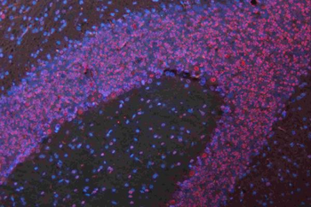
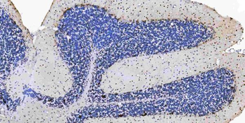
IF analysis of Musashi 1/Msi1 using anti-Musashi 1/Msi1 antibody. Musashi 1/Msi1 was detected in a paraffin-embedded section of rat cerebellum tissue. Heat mediated antigen retrieval was performed in EDTA buffer (pH8.0, epitope retrieval solution). The tissue section was blocked with 10% goat serum. The tissue section was then incubated with 25 µg/mL rabbit anti-Musashi 1/Msi1 Antibody overnight at 4°C. DyLight®594 Conjugated Goat Anti-Rabbit IgG was used as secondary antibody at 1:100 dilution and incubated for 30 minutes at 37°C. The section was counterstained with DAPI. Visualize using a fluorescence microscope and filter sets appropriate for the label used.
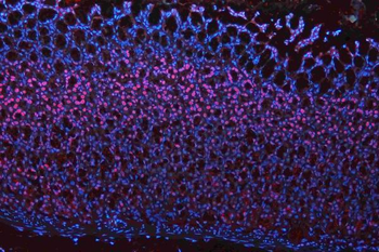
IF analysis of Musashi 1/Msi1 using anti-Musashi 1/Msi1 antibody. Musashi 1/Msi1 was detected in a paraffin-embedded section of rat stomach tissue. Heat mediated antigen retrieval was performed in EDTA buffer (pH8.0, epitope retrieval solution). The tissue section was blocked with 10% goat serum. The tissue section was then incubated with 25 µg/mL rabbit anti-Musashi 1/Msi1 Antibody overnight at 4°C. DyLight®594 Conjugated Goat Anti-Rabbit IgG was used as secondary antibody at 1:100 dilution and incubated for 30 minutes at 37°C. The section was counterstained with DAPI. Visualize using a fluorescence microscope and filter sets appropriate for the label used.
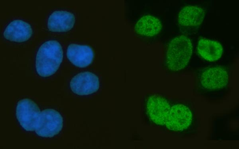
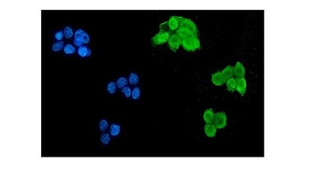
IF analysis of Musashi 1/Msi1 using anti-Musashi 1/Msi1 antibody. Musashi 1/Msi1 was detected in an immunocytochemical section of MCF7 cells. Enzyme antigen retrieval was performed using IHC enzyme antigen retrieval reagent for 15 mins. The cells were blocked with 10% goat serum. And then incubated with 5 µg/mL rabbit anti-Musashi 1/Msi1 Antibody overnight at 4°C. DyLight®488 Conjugated Goat Anti-Rabbit IgG was used as secondary antibody at 1:500 dilution and incubated for 30 minutes at 37°C. The section was counterstained with DAPI. Visualize using a fluorescence microscope and filter sets appropriate for the label used.
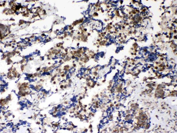
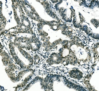
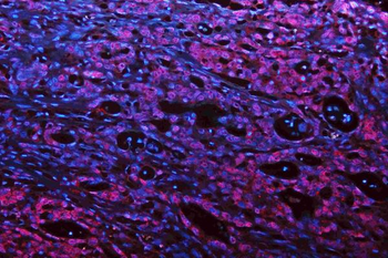
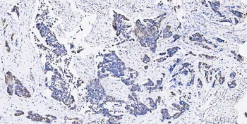
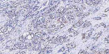
IHC analysis of Musashi 1/Msi1 using anti-Musashi 1/Msi1 antibody. Musashi 1/Msi1 was detected in paraffin-embedded section of human lung cancer tissues. Heat mediated antigen retrieval was performed in citrate buffer (pH6, epitope retrieval solution) for 20 mins. The tissue section was blocked with 10% goat serum. The tissue section was then incubated with 1 µg/ml rabbit anti-Musashi 1/Msi1 Antibody overnight at 4°C. Biotinylated goat anti-rabbit IgG was used as secondary antibody and incubated for 30 minutes at 37°C. The tissue section was developed using Strepavidin-Biotin-Complex (SABC) with DAB as the chromogen.
IHC analysis of Musashi 1/Msi1 using anti-Musashi 1/Msi1 antibody. Musashi 1/Msi1 was detected in paraffin-embedded section of human mammary cancer tissues. Heat mediated antigen retrieval was performed in citrate buffer (pH6, epitope retrieval solution) for 20 mins. The tissue section was blocked with 10% goat serum. The tissue section was then incubated with 1 µg/ml rabbit anti-Musashi 1/Msi1 Antibody overnight at 4°C. Biotinylated goat anti-rabbit IgG was used as secondary antibody and incubated for 30 minutes at 37°C. The tissue section was developed using Strepavidin-Biotin-Complex (SABC) with DAB as the chromogen.
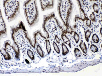
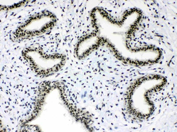
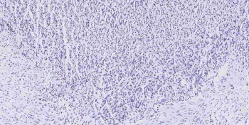
IHC analysis of Musashi 1/Msi1 using anti-Musashi 1/Msi1 antibody. Musashi 1/Msi1 was detected in paraffin-embedded section of mouse intestine tissues. Heat mediated antigen retrieval was performed in citrate buffer (pH6, epitope retrieval solution) for 20 mins. The tissue section was blocked with 10% goat serum. The tissue section was then incubated with 1 µg/ml rabbit anti-Musashi 1/Msi1 Antibody overnight at 4°C. Biotinylated goat anti-rabbit IgG was used as secondary antibody and incubated for 30 minutes at 37°C. The tissue section was developed using Strepavidin-Biotin-Complex (SABC) with DAB as the chromogen.
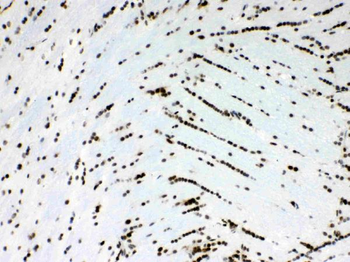
IHC analysis of Musashi 1/Msi1 using anti-Musashi 1/Msi1 antibody. Musashi 1/Msi1 was detected in paraffin-embedded section of rat brain tissues. Heat mediated antigen retrieval was performed in citrate buffer (pH6, epitope retrieval solution) for 20 mins. The tissue section was blocked with 10% goat serum. The tissue section was then incubated with 1 µg/ml rabbit anti-Musashi 1/Msi1 Antibody overnight at 4°C. Biotinylated goat anti-rabbit IgG was used as secondary antibody and incubated for 30 minutes at 37°C. The tissue section was developed using Strepavidin-Biotin-Complex (SABC) with DAB as the chromogen.
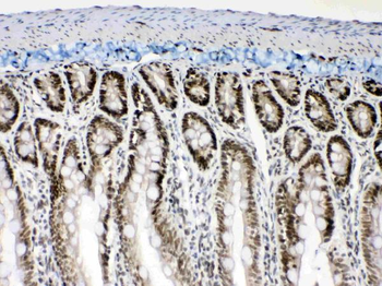
IHC analysis of Musashi 1/Msi1 using anti-Musashi 1/Msi1 antibody. Musashi 1/Msi1 was detected in paraffin-embedded section of rat intestine tissues. Heat mediated antigen retrieval was performed in citrate buffer (pH6, epitope retrieval solution) for 20 mins. The tissue section was blocked with 10% goat serum. The tissue section was then incubated with 1 µg/ml rabbit anti-Musashi 1/Msi1 Antibody overnight at 4°C. Biotinylated goat anti-rabbit IgG was used as secondary antibody and incubated for 30 minutes at 37°C. The tissue section was developed using Strepavidin-Biotin-Complex (SABC) with DAB as the chromogen.
IHC analysis of Musashi 1/Msi1 using anti-Musashi 1/Msi1 antibody. Musashi 1/Msi1 was detected in a paraffin-embedded section of human breast cancer tissue. Heat mediated antigen retrieval was performed in EDTA buffer (pH8.0, epitope retrieval solution). The tissue section was blocked with 10% goat serum. The tissue section was then incubated with 2.5 µg/ml rabbit anti-Musashi 1/Msi1 Antibody overnight at 4°C. HRP Conjugated Goat Anti-rabbit IgG was used as secondary antibody and incubated for 30 minutes at 37°C. The tissue section was developed using HRP Conjugated Rabbit IgG Super Vision Assay Kit with DAB as the chromogen.
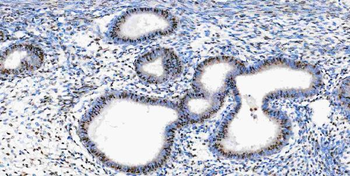
IHC analysis of Musashi 1/Msi1 using anti-Musashi 1/Msi1 antibody. Musashi 1/Msi1 was detected in a paraffin-embedded section of human endometrial cancer tissue. Heat mediated antigen retrieval was performed in EDTA buffer (pH8.0, epitope retrieval solution). The tissue section was blocked with 10% goat serum. The tissue section was then incubated with 2.5 µg/ml rabbit anti-Musashi 1/Msi1 Antibody overnight at 4°C. HRP Conjugated Goat Anti-rabbit IgG was used as secondary antibody and incubated for 30 minutes at 37°C. The tissue section was developed using HRP Conjugated Rabbit IgG Super Vision Assay Kit with DAB as the chromogen.
IHC analysis of Musashi 1/Msi1 using anti-Musashi 1/Msi1 antibody. Musashi 1/Msi1 was detected in a paraffin-embedded section of human ovarian cancer tissue. Heat mediated antigen retrieval was performed in EDTA buffer (pH8.0, epitope retrieval solution). The tissue section was blocked with 10% goat serum. The tissue section was then incubated with 2.5 µg/ml rabbit anti-Musashi 1/Msi1 Antibody overnight at 4°C. HRP Conjugated Goat Anti-rabbit IgG was used as secondary antibody and incubated for 30 minutes at 37°C. The tissue section was developed using HRP Conjugated Rabbit IgG Super Vision Assay Kit with DAB as the chromogen.
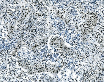
IHC analysis of Musashi 1/Msi1 using anti-Musashi 1/Msi1 antibody. Musashi 1/Msi1 was detected in a paraffin-embedded section of human pancreatic cancer tissue. Heat mediated antigen retrieval was performed in EDTA buffer (pH8.0, epitope retrieval solution). The tissue section was blocked with 10% goat serum. The tissue section was then incubated with 2.5 µg/ml rabbit anti-Musashi 1/Msi1 Antibody overnight at 4°C. HRP Conjugated Goat Anti-rabbit IgG was used as secondary antibody and incubated for 30 minutes at 37°C. The tissue section was developed using HRP Conjugated Rabbit IgG Super Vision Assay Kit with DAB as the chromogen.
IHC analysis of Musashi 1/Msi1 using anti-Musashi 1/Msi1 antibody. Musashi 1/Msi1 was detected in a paraffin-embedded section of human pancreatic cancer tissue. Heat mediated antigen retrieval was performed in EDTA buffer (pH8.0, epitope retrieval solution). The tissue section was blocked with 10% goat serum. The tissue section was then incubated with 2.5 µg/ml rabbit anti-Musashi 1/Msi1 Antibody overnight at 4°C. HRP Conjugated Goat Anti-rabbit IgG was used as secondary antibody and incubated for 30 minutes at 37°C. The tissue section was developed using HRP Conjugated Rabbit IgG Super Vision Assay Kit with DAB as the chromogen.
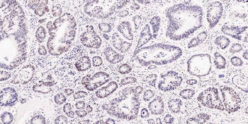
IHC analysis of Musashi 1/Msi1 using anti-Musashi 1/Msi1 antibody. Musashi 1/Msi1 was detected in a paraffin-embedded section of human stomach cancer tissue. Heat mediated antigen retrieval was performed in EDTA buffer (pH8.0, epitope retrieval solution). The tissue section was blocked with 10% goat serum. The tissue section was then incubated with 2.5 µg/ml rabbit anti-Musashi 1/Msi1 Antibody overnight at 4°C. HRP Conjugated Goat Anti-rabbit IgG was used as secondary antibody and incubated for 30 minutes at 37°C. The tissue section was developed using HRP Conjugated Rabbit IgG Super Vision Assay Kit with DAB as the chromogen.
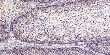
IHC analysis of Musashi 1/Msi1 using anti-Musashi 1/Msi1 antibody. Musashi 1/Msi1 was detected in a paraffin-embedded section of mouse brain tissue. Heat mediated antigen retrieval was performed in EDTA buffer (pH8.0, epitope retrieval solution). The tissue section was blocked with 10% goat serum. The tissue section was then incubated with 2.5 µg/ml rabbit anti-Musashi 1/Msi1 Antibody overnight at 4°C. HRP Conjugated Goat Anti-rabbit IgG was used as secondary antibody and incubated for 30 minutes at 37°C. The tissue section was developed using HRP Conjugated Rabbit IgG Super Vision Assay Kit with DAB as the chromogen.
IHC analysis of Musashi 1/Msi1 using anti-Musashi 1/Msi1 antibody. Musashi 1/Msi1 was detected in a paraffin-embedded section of mouse cerebellum tissue. Heat mediated antigen retrieval was performed in EDTA buffer (pH8.0, epitope retrieval solution). The tissue section was blocked with 10% goat serum. The tissue section was then incubated with 2.5 µg/ml rabbit anti-Musashi 1/Msi1 Antibody overnight at 4°C. HRP Conjugated Goat Anti-rabbit IgG was used as secondary antibody and incubated for 30 minutes at 37°C. The tissue section was developed using HRP Conjugated Rabbit IgG Super Vision Assay Kit with DAB as the chromogen.
IHC analysis of Musashi 1/Msi1 using anti-Musashi 1/Msi1 antibody. Musashi 1/Msi1 was detected in a paraffin-embedded section of mouse stomach tissue. Heat mediated antigen retrieval was performed in EDTA buffer (pH8.0, epitope retrieval solution). The tissue section was blocked with 10% goat serum. The tissue section was then incubated with 2.5 µg/ml rabbit anti-Musashi 1/Msi1 Antibody overnight at 4°C. HRP Conjugated Goat Anti-rabbit IgG was used as secondary antibody and incubated for 30 minutes at 37°C. The tissue section was developed using HRP Conjugated Rabbit IgG Super Vision Assay Kit with DAB as the chromogen.
IHC analysis of Musashi 1/Msi1 using anti-Musashi 1/Msi1 antibody. Musashi 1/Msi1 was detected in a paraffin-embedded section of rat cerebellum tissue. Heat mediated antigen retrieval was performed in EDTA buffer (pH8.0, epitope retrieval solution). The tissue section was blocked with 10% goat serum. The tissue section was then incubated with 2.5 µg/ml rabbit anti-Musashi 1/Msi1 Antibody overnight at 4°C. HRP Conjugated Goat Anti-rabbit IgG was used as secondary antibody and incubated for 30 minutes at 37°C. The tissue section was developed using HRP Conjugated Rabbit IgG Super Vision Assay Kit with DAB as the chromogen.
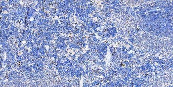
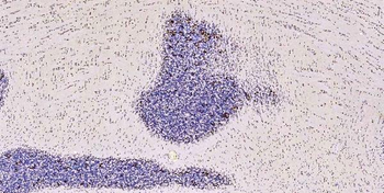
IHC analysis of Musashi 1/Msi1 using anti-Musashi 1/Msi1 antibody. Musashi 1/Msi1 was detected in a paraffin-embedded section of rat spleen tissue. Heat mediated antigen retrieval was performed in EDTA buffer (pH8.0, epitope retrieval solution). The tissue section was blocked with 10% goat serum. The tissue section was then incubated with 2.5 µg/ml rabbit anti-Musashi 1/Msi1 Antibody overnight at 4°C. HRP Conjugated Goat Anti-rabbit IgG was used as secondary antibody and incubated for 30 minutes at 37°C. The tissue section was developed using HRP Conjugated Rabbit IgG Super Vision Assay Kit with DAB as the chromogen.
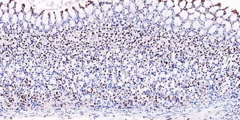
IHC analysis of Musashi 1/Msi1 using anti-Musashi 1/Msi1 antibody. Musashi 1/Msi1 was detected in a paraffin-embedded section of rat stomach tissue. Heat mediated antigen retrieval was performed in EDTA buffer (pH8.0, epitope retrieval solution). The tissue section was blocked with 10% goat serum. The tissue section was then incubated with 2.5 µg/ml rabbit anti-Musashi 1/Msi1 Antibody overnight at 4°C. HRP Conjugated Goat Anti-rabbit IgG was used as secondary antibody and incubated for 30 minutes at 37°C. The tissue section was developed using HRP Conjugated Rabbit IgG Super Vision Assay Kit with DAB as the chromogen.
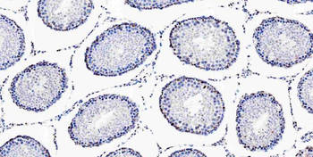
IHC analysis of Musashi 1/Msi1 using anti-Musashi 1/Msi1 antibody. Musashi 1/Msi1 was detected in a paraffin-embedded section of rat testis tissue. Heat mediated antigen retrieval was performed in EDTA buffer (pH8.0, epitope retrieval solution). The tissue section was blocked with 10% goat serum. The tissue section was then incubated with 2.5 µg/ml rabbit anti-Musashi 1/Msi1 Antibody overnight at 4°C. HRP Conjugated Goat Anti-rabbit IgG was used as secondary antibody and incubated for 30 minutes at 37°C. The tissue section was developed using HRP Conjugated Rabbit IgG Super Vision Assay Kit with DAB as the chromogen.
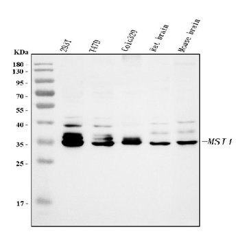
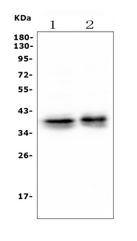
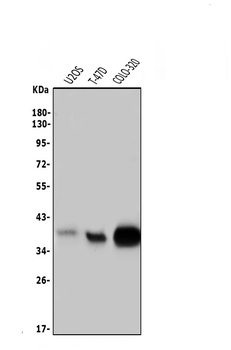
Western blot analysis of Musashi 1/Msi1 using anti-Musashi 1/Msi1 antibody. Electrophoresis was performed on a 5-20% SDS-PAGE gel at 70V (Stacking gel) / 90V (Resolving gel) for 2-3 hours. The sample well of each lane was loaded with 30 ug of sample under reducing conditions. Lane 1: human 293T whole cell lysates, Lane 2: human T-47D whole cell lysates, Lane 3: human Colo320 whole cell lysates, Lane 4: rat brain tissue lysates, Lane 5: mouse brain tissue lysates. After electrophoresis, proteins were transferred to a nitrocellulose membrane at 150 mA for 50-90 minutes. Blocked the membrane with 5% non-fat milk/TBS for 1.5 hour at RT. The membrane was incubated with rabbit anti-Musashi 1/Msi1 antigen affinity purified polyclonal antibody at 0.5 µg/mL overnight at 4°C, then washed with TBS-0.1% Tween 3 times with 5 minutes each and probed with a goat anti-rabbit IgG-HRP secondary antibody at a dilution of 1:5000 for 1.5 hour at RT. The signal is developed using an Enhanced Chemiluminescent detection (ECL) kit with Tanon 5200 system. A specific band was detected for Musashi 1/Msi1 at approximately 39 kDa. The expected band size for Musashi 1/Msi1 is at 39 kDa.
- Item 1 of 4
Anti-Musashi 1/Msi1 Antibody Picoband (monoclonal, 2B9) [orb623779]
ICC, IF, IHC, WB
Human, Mouse, Rat
Mouse
Monoclonal
Unconjugated
10 μg, 100 μg - Item 1 of 2
Anti-Musashi 1/Msi1 Antibody Picoband [orb654349]
ELISA, FC, WB
Human
Rabbit
Polyclonal
Unconjugated
10 μg, 100 μg 
Anti-Musashi 1/Msi1 Antibody Picoband [orb2606316]
ELISA, FC, WB
Human
Rabbit
Polyclonal
iFluor647
100 μg