You have no items in your shopping cart.
Cart summary
Item 1 of 3
Item 1 of 3
Anti-Hu CD54 Purified Azide Free
Catalog Number: orb43579

| Catalog Number | orb43579 |
|---|---|
| Category | Antibodies |
| Description | Mouse Monoclonal to CD54. |
| Clonality | Monoclonal |
| Clone Number | 1H4 |
| Tested applications | FC, ICC, IHC-Fr, IHC-P, WB |
| Reactivity | Human |
| Isotype | Mouse IgG2b |
| Immunogen | Raji cells and spleen cells fused with NS1 cells |
| Antibody Type | Primary Antibody |
| Concentration | 1 mg/ml |
| Dilution range | Immunohistochemistry (frozen sections): Recommended dilution: 5-10 μg/ml. Immunohistochemistry (paraffin sections): Recommended dilution: 10 μg/ml; prolonged fixation in buffered formalin can destroy the epitope. High temperature antigen unmasking technique is required. Flow cytometry: Recommended dilution: 1-4 μg/ml. Western blotting: Recommended dilution: 1-2 μg/ml; non-reducing conditions. |
| Purity | Purified by protein-A affinity chromatography. |
| Conjugation | Unconjugated |
| Target | CD54 |
| Entrez | 3383 |
| UniProt ID | P05362 |
| RRID | AB_10995858 |
| Storage | Maintain refrigerated at 2-8°C for up to 2 weeks. For long term storage store at -20°C in small aliquots to prevent freeze-thaw cycles. |
| Buffer/Preservatives | Phosphate buffered saline (PBS), pH 7.4 |
| Alternative names | Anti-CD54 antibody, anti-ICAM-1 antibody Read more... |
| Note | For research use only |
| Application notes | Immunohistochemistry (frozen sections): Recommended dilution: 5-10 μg/ml. Immunohistochemistry (paraffin sections): Recommended dilution: 10 μg/ml; prolonged fixation in buffered formalin can destroy the epitope. High temperature antigen unmasking technique is required. Flow cytometry: Recommended dilution: 1-4 μg/ml.Western blotting: Recommended dilution: 1-2 μg/ml; non-reducing conditions. |
| Expiration Date | 12 months from date of receipt. |

Flow cytometry surface staining pattern of human peripheral whole blood stained using anti-human CD54 (1H4) purified antibody (concentration in sample 3 μg/ml, GAM FITC).
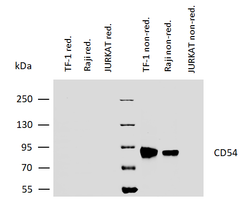
Western blotting analysis of human CD54 using mouse monoclonal antibody 1H4 on lysates of TF-1 and Raji cells, as well as of JURKAT cells (negative control) under reducing and non-reducing conditions. Nitrocellulose membrane was probed with 2 µg/ml of mouse anti-CD54 monoclonal antibody followed by IRDye800-conjugated anti-mouse secondary antibody. A specific band was detected for CD54 at approximately 88 kDa.
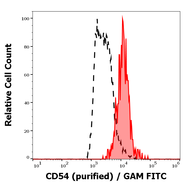
Separation of human monocytes (red-filled) from human lymphocytes (black-dashed) in flow cytometry analysis (surface staining) of peripheral whole blood stained using anti-human CD54 (1H4) purified antibody (concentration in sample 3 μg/ml, GAM FITC).
