You have no items in your shopping cart.
Cart summary
Item 1 of 7
Item 1 of 7
Anti-Hu CD16 FITC
Catalog Number: orb44655

| Catalog Number | orb44655 |
|---|---|
| Category | Antibodies |
| Description | Mouse Monoclonal to CD16. |
| Clonality | Monoclonal |
| Clone Number | 3G8 |
| Tested applications | FC |
| Reactivity | Human, Primate |
| Isotype | Mouse IgG1 kappa |
| Immunogen | Human neutrophils |
| Antibody Type | Primary Antibody |
| Purity | Purified antibody is conjugated with fluorescein isothiocyanate (FITC) under optimum conditions and unconjugated antibody and free fluorochrome are removed by size-exclusion chromatography. |
| Conjugation | FITC |
| Target | CD16 |
| RRID | AB_10993896 |
| Storage | Store at 2-8°C. Protect from prolonged exposure to light. Do not freeze. |
| Buffer/Preservatives | Stabilizing phosphate buffered saline (PBS), pH 7.4, 15 mM sodium azide |
| Alternative names | Anti-CD16 antibody, anti-FACSgammaRIII antibody Read more... |
| Note | For research use only |
| Application notes | Flow cytometry: The reagent is designed for analysis of human blood cells using 4 μl reagent / 100 μl of whole blood or 106 cells in a suspension. The content of a vial (0.4 ml) is sufficient for 100 tests. |
| Expiration Date | 12 months from date of receipt. |
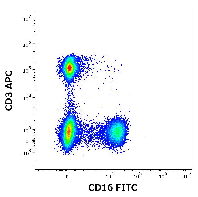
Staining pattern of Anti-human CD16 FITC antibody (clone 3G8) in dot-plot fluorescence visualization (lymphocyte gate) Analysis of the antibody staining profile was performed on blood leukocytes isolated from peripheral whole blood by bulk erythrocyte lysis using 10× diluted EXCELLYSE Live. Mouse monoclonal anti-human CD16 FITC antibody (clone 3G8) was used in concentration 9 µg/ml in stained stained blood sample (2 x 10^6 cells) and Mouse monoclonal anti-human CD3 APC antibody (clone UCHT1) in concentration 6 µg/ml, respectively.
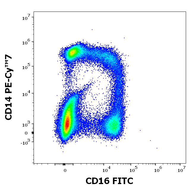
Staining pattern of Anti-human CD16 FITC antibody (clone 3G8) in dot-plot fluorescence visualization (mononuclear gate) Analysis of the antibody staining profile was performed on blood leukocytes isolated from peripheral whole blood by bulk erythrocyte lysis using 10× diluted EXCELLYSE Live. Mouse monoclonal anti-human CD16 FITC antibody (clone 3G8) was used in concentration 9 µg/ml in stained stained blood sample (2 x 10^6 cells) and Mouse monoclonal anti-human CD14 PE-Cy™7 antibody (clone MEM-15) in concentration 8 µg/ml, respectively.
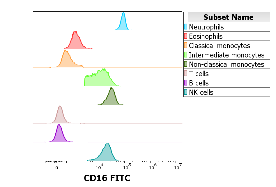
Reactivity of Anti-human CD16 FITC antibody (clone 3G8) on human peripheral leukocytes Analysis of the antibody staining profile was performed on blood leukocytes isolated from peripheral whole blood by bulk erythrocyte lysis using 10× diluted EXCELLYSE Live. Suspension of blood leukocytes (2 x 10^6 cells) was added to the mixture of CD16 FITC antibody (clone 3G8, 9 µg/ml in stained blood sample), backbone antibody conjugates and Monocyte Blocking Buffer, vortexed and incubated for 20 min. Stained sample was fixed with 2 ml of 10× diluted EXCELLYSE Easy solution for 10 min. Finally, samples were centrifuged (670 g, 5 min.), supernatant removed and the cell pellet was resuspended in 200 µl of PBS for acquisition.
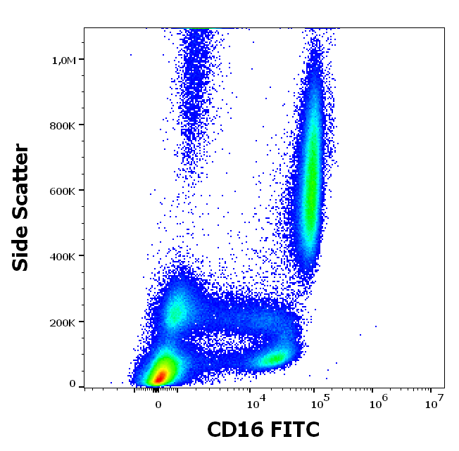
Anti-human CD16 FITC antibody (clone 3G8) works in flow cytometry application. Analysis of the antibody staining profile was performed on blood leukocytes isolated from peripheral whole blood by bulk erythrocyte lysis using 10× diluted EXCELLYSE Live. Mouse monoclonal anti-human CD16 FITC antibody (clone 3G8) was used in concentration 9 µg/ml in stained blood sample (2 x 10^6 cells).
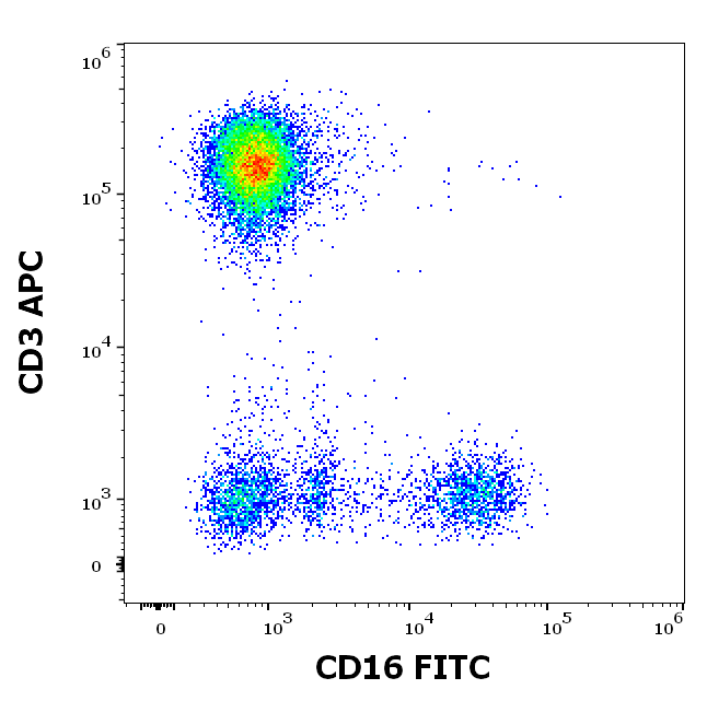
Flow cytometry multicolor surface staining pattern of human peripheral whole blood stained using anti-human CD16 (3G8) FITC antibody (4 μl reagent / 100 μl of peripheral whole blood) and anti-human CD3 (UCHT1) APC antibody (10 μl reagent / 100 μl of peripheral whole blood).
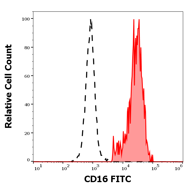
Separation of human CD16 positive CD3 negative lymphocytes (red-filled) from CD16 negative CD3 positive lymphocytes (black-dashed) in flow cytometry analysis (surface staining) of human peripheral whole blood stained using anti-human CD16 (3G8) FITC antibody (4 μl reagent / 100 μl of peripheral whole blood).
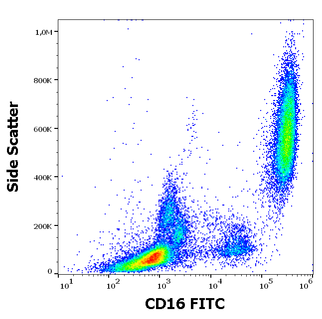
Flow cytometry surface staining pattern of human peripheral whole blood stained using anti-human CD16 (3G8) FITC antibody (4 μl reagent / 100 μl of peripheral whole blood).
- Item 1 of 4
- Item 1 of 2


