You have no items in your shopping cart.
Cart summary
Item 1 of 2
Item 1 of 2
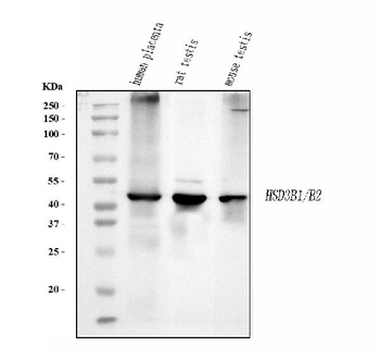
Anti-HSD3B1 Antibody
Catalog Number: orb412672

| Catalog Number | orb412672 |
|---|---|
| Category | Antibodies |
| Description | Rabbit polyclonal antibody to HSD3B1 |
| Species/Host | Rabbit |
| Clonality | Polyclonal |
| Tested applications | IF, WB |
| Reactivity | Human, Mouse, Rat |
| Immunogen | Recombinant full length protein of human HSD3B1 |
| Dilution range | WB: 1:500-2000 |
| Form/Appearance | Liquid in 0.42% Potassium phosphate, 0.87% Sodium chloride, pH 7.3, 30% glycerol, and 0.01% sodium azide. |
| Conjugation | Unconjugated |
| Target | HSD3B1 |
| Entrez | 15492, 3283, 360348 |
| UniProt ID | P22071, P24815, P14060 |
| Source | Rabbit |
| Storage | Maintain refrigerated at 2-8°C for up to 2 weeks. For long term storage store at -20°C in small aliquots to prevent freeze-thaw cycles. |
| Buffer/Preservatives | Liquid in 0.42% Potassium phosphate, 0.87% Sodium chloride, pH 7.3, 30% glycerol, and 0.01% sodium azide. |
| Alternative names | anti-3BH antibody, anti-HSDB3A antibody, anti-3 be Read more... |
| Note | For research use only |
| Expiration Date | 12 months from date of receipt. |

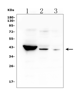
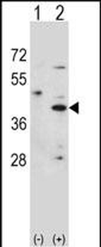
Western blot analysis of HSD3B1 expression in A375 (A) whole cell lysates. (Predicted band size: 42 kD; Observed band size: 42 kD)

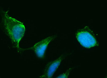
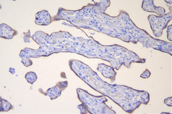
Immunofluorescent analysis of HSD3B1 staining in human placenta. Formalin-fixed cells were permeabilized with 0.1% Triton X-100 in TBS for 5-10 minutes and blocked with 3% BSA-PBS for 30 minutes at room temperature. Cells were probed with the primary antibody in 3% BSA-PBS and incubated overnight at 4 °C in a humidified chamber. Cells were washed with PBST and incubated with a AF594-conjugated secondary antibody (red) in PBS at room temperature in the dark. DAPI was used to stain the cell nuclei (blue).
- Item 1 of 4
- Item 1 of 4
Anti-HSD3B1 Antibody [orb614122]
ELISA, FC, ICC, IF, IHC, WB
Human
Rabbit
Polyclonal
Unconjugated
10 μg, 100 μg - Item 1 of 3
- Item 1 of 2
Anti-HSD3B1 Antibody (monoclonal, 3F3C/7F9C8) [orb865663]
ICC, IF, WB
Human, Mouse, Rat
Mouse
Monoclonal
Unconjugated
100 μg, 10 μg - Item 1 of 1
Anti-HSD3B1 Rabbit Monoclonal Antibody [orb866665]
ICC, IF, WB
Human
Rabbit
Monoclonal
Unconjugated
30 μl, 100 μl