You have no items in your shopping cart.
Cart summary
Item 1 of 5
Item 1 of 5
Anti-Fos B/FOSB Antibody
Catalog Number: orb763044

| Catalog Number | orb763044 |
|---|---|
| Category | Antibodies |
| Description | Anti-Fos B/FOSB Antibody. Tested in ELISA, IF, IHC, ICC applications. This antibody reacts with Human. |
| Species/Host | Rabbit |
| Clonality | Polyclonal |
| Tested applications | ELISA, ICC, IF, IHC |
| Reactivity | Human |
| Isotype | Rabbit IgG |
| Immunogen | E.coli-derived human FOSB recombinant protein (Position: M1-S332). |
| Antibody Type | Primary Antibody |
| Concentration | Adding 0.2 ml of distilled water will yield a concentration of 500 μg/ml. |
| Form/Appearance | Lyophilized |
| Conjugation | Unconjugated |
| MW | 75 kDa |
| UniProt ID | P53539 |
| Storage | Maintain refrigerated at 2-8°C for up to 2 weeks. For long term storage store at -20°C in small aliquots to prevent freeze-thaw cycles. |
| Alternative names | Vitamin K-dependent protein S; PROS1; PROS Read more... |
| Note | For research use only |
| Application notes | Immunohistochemistry (Paraffin-embedded Section), 2-5μg/ml, Human Immunocytochemistry/Immunofluorescence, 5μg/ml, Human ELISA, 0.1-0.5μg/ml, -. Add 0.2ml of distilled water will yield a concentration of 500ug/ml |
| Expiration Date | 12 months from date of receipt. |

IF analysis of Fos B/FOSB using anti-Fos B/FOSB antibody. Fos B/FOSB was detected in an immunocytochemical section of SiHa cells. Enzyme antigen retrieval was performed using IHC enzyme antigen retrieval reagent for 15 mins. The cells were blocked with 10% goat serum. And then incubated with 5 µg/mL rabbit anti-Fos B/FOSB Antibody overnight at 4°C. DyLight®488 Conjugated Goat Anti-Rabbit IgG was used as secondary antibody at 1:100 dilution and incubated for 30 minutes at 37°C. The section was counterstained with DAPI. Visualize using a fluorescence microscope and filter sets appropriate for the label used.
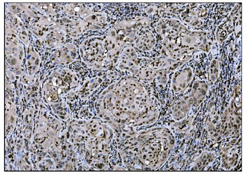
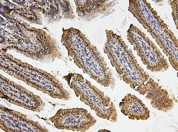
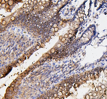
IHC analysis of Fos B/FOSB using anti-Fos B/FOSB antibody. Fos B/FOSB was detected in a paraffin-embedded section of human gallbladder adenocarcinoma tissue. Heat mediated antigen retrieval was performed in EDTA buffer (pH8.0, epitope retrieval solution). The tissue section was blocked with 10% goat serum. The tissue section was then incubated with 2 µg/ml rabbit anti-Fos B/FOSB Antibody overnight at 4°C. Biotinylated goat anti-rabbit IgG was used as secondary antibody and incubated for 30 minutes at 37°C. The tissue section was developed using Strepavidin-Biotin-Complex (SABC) with DAB as the chromogen.
IHC analysis of Fos B/FOSB using anti-Fos B/FOSB antibody. Fos B/FOSB was detected in a paraffin-embedded section of human gallbladder adenocarcinoma tissue. Heat mediated antigen retrieval was performed in EDTA buffer (pH8.0, epitope retrieval solution). The tissue section was blocked with 10% goat serum. The tissue section was then incubated with 2 µg/ml rabbit anti-Fos B/FOSB Antibody overnight at 4°C. Biotinylated goat anti-rabbit IgG was used as secondary antibody and incubated for 30 minutes at 37°C. The tissue section was developed using Strepavidin-Biotin-Complex (SABC) with DAB as the chromogen.
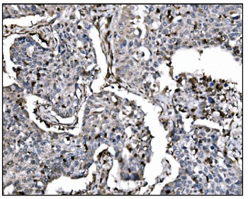
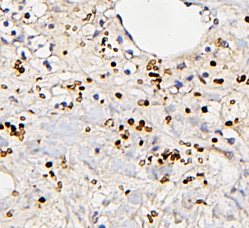
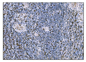
IHC analysis of Fos B/FOSB using anti-Fos B/FOSB antibody. Fos B/FOSB was detected in a paraffin-embedded section of human lung cancer tissue. Heat mediated antigen retrieval was performed in EDTA buffer (pH8.0, epitope retrieval solution). The tissue section was blocked with 10% goat serum. The tissue section was then incubated with 2 µg/ml rabbit anti-Fos B/FOSB Antibody overnight at 4°C. Biotinylated goat anti-rabbit IgG was used as secondary antibody and incubated for 30 minutes at 37°C. The tissue section was developed using Strepavidin-Biotin-Complex (SABC) with DAB as the chromogen.
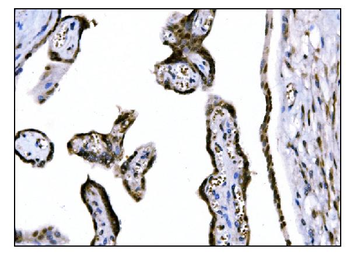
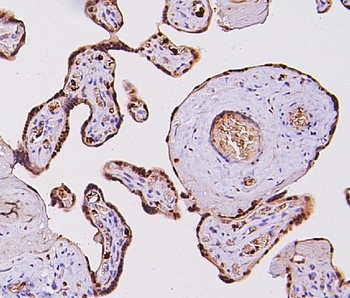
IHC analysis of Fos B/FOSB using anti-Fos B/FOSB antibody. Fos B/FOSB was detected in a paraffin-embedded section of human placenta tissue. Heat mediated antigen retrieval was performed in EDTA buffer (pH8.0, epitope retrieval solution). The tissue section was blocked with 10% goat serum. The tissue section was then incubated with 2 µg/ml rabbit anti-Fos B/FOSB Antibody overnight at 4°C. Biotinylated goat anti-rabbit IgG was used as secondary antibody and incubated for 30 minutes at 37°C. The tissue section was developed using Strepavidin-Biotin-Complex (SABC) with DAB as the chromogen.
- Item 1 of 6
Anti-Fos B/FOSB Antibody [orb312091]
FC, IHC, WB
Human, Mouse, Rat
Rabbit
Polyclonal
Unconjugated
100 μg, 10 μg - Item 1 of 1