You have no items in your shopping cart.
Cart summary
Item 1 of 6
Item 1 of 6
Anti-Cytokeratins Purified
Catalog Number: orb43707

| Catalog Number | orb43707 |
|---|---|
| Category | Antibodies |
| Description | Mouse monoclonal antibody to pan Cytokeratin |
| Clonality | Monoclonal |
| Clone Number | C-11 |
| Tested applications | FC, ICC, IHC-P, IP, WB |
| Reactivity | Mammal |
| Isotype | Mouse IgG1 |
| Immunogen | Keratin-enriched preparation from human epidermoid carcinoma cell line A431. |
| Concentration | 1 mg/ml |
| Purity | Purified by protein-A affinity chromatography. |
| Conjugation | Unconjugated |
| Target | Cytokeratins |
| RRID | AB_10992679 |
| Storage | Maintain refrigerated at 2-8°C for up to 2 weeks. For long term storage store at -20°C in small aliquots to prevent freeze-thaw cycles. |
| Buffer/Preservatives | Phosphate buffered saline (PBS), pH 7.4, 15 mM sodium azide |
| Alternative names | anti pan-cytokeratin antibody, anti pan-CK antibod Read more... |
| Note | For research use only |
| Application notes | Flow cytometry: Recommended dilution: 1 μg/ml. Intracellular staining.Immunohistochemistry: Recommended dilution: 2-8 μg/ml.Western blotting: Recommended dilution: 1-2 μg/ml. |
| Expiration Date | 12 months from date of receipt. |
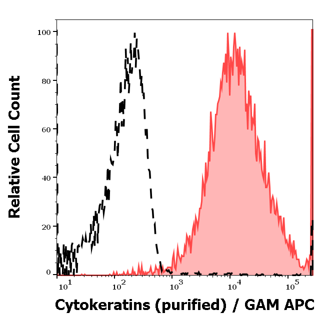
Separation of MCF-7 cells (red-filled) from human leukocytes (black-dashed) in flow cytometry analysis (intracellular staining) of peripheral whole blood spiked with MCF-7 cells stained using anti-Cytokeratins (C-11) purified antibody (concentration in sample 3 µg/ml, GAM APC).

Flow cytometry intracellular staining pattern of human peripheral whole blood spiked with MCF-7 cells stained using anti-Cytokeratins (C-11) purified antibody (concentration in sample 3 µg/ml, GAM APC).
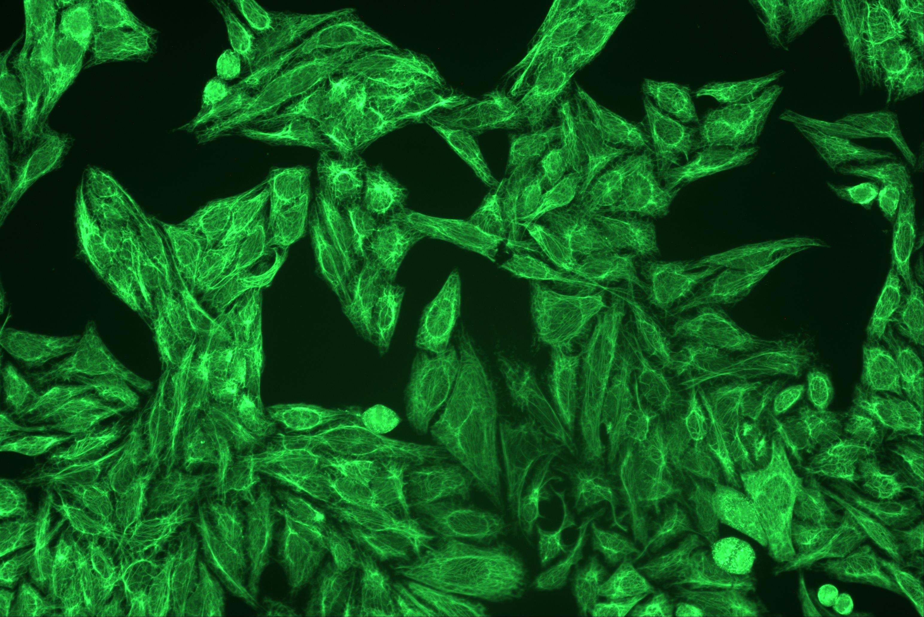
Immunocytochemistry staining of cytokeratins in Hep-2 cells using pan-cytokeratin antibody C-11 (orb43707, diluted 1:400), detected with GAM IgG-Alexa Fluor®488 (diluted 1:200; green).
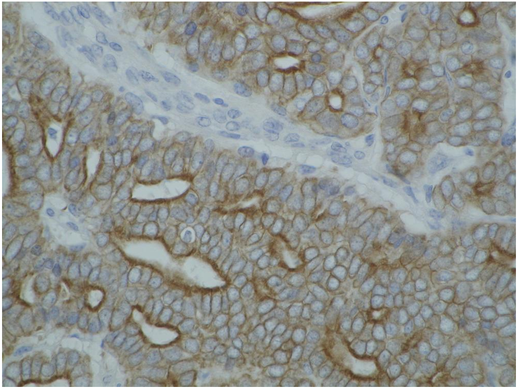
Immunohistochemistry staining of cytokeratin on paraffin-embedded sections of guinea pig breast carcinoma using anti-cytokeratin antibody (C-11).
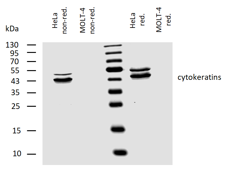
Western blotting analysis of human cytokeratins using mouse monoclonal antibody C-11 on lysates of HeLa cell line and MOLT-4 cell line (cytokeratin non-expressing cell line; negative control) under non-reducing and reducing conditions. Nitrocellulose membrane was probed with 2 µg/ml of mouse monoclonal antibody anti-cytokeratins followed by IRDye800-conjugated anti-mouse secondary antibody. Specific bands were detected for cytokeratins at approximately 45-55 kDa.
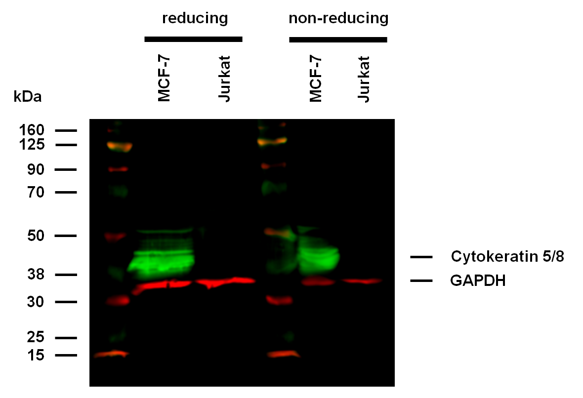
Anti-Hu Cytokeratin 5/8 (clone C-50) works in WB application under reducing and non-reducing conditions. Western blotting analysis was performed on whole cell extracts (RIPA lysis buffer) of MCF-7 and Jurkat cell lines, mixed and heated (100°C, 5 min) with reducing and non-reducing SDS-loading buffer. Samples were resolved using 10% Tris-glycine SDS gel electrophoresis. Nitrocellulose membrane blot was probed with mouse IgG1 monoclonal antibody C-50 (1 µg/ml), followed by IRDye 800CW Goat-anti-Mouse IgG (green). Mouse anti-GAPDH monoclonal antibody FF26A conjugated with DyLight 680 (0.1 µg/ml), was used as the loading control (red). Multiplex fluorescent Western blot detection was performed. Cytokeratin 5/8 molecules were detected at ~40 kDa in MCF-7 cell line under both reducing and non-reducing conditions.
