You have no items in your shopping cart.
Cart summary
Item 1 of 16
Item 1 of 16
Anti-Cytokeratin 8/KRT8 Antibody
Catalog Number: orb19070

| Catalog Number | orb19070 |
|---|---|
| Category | Antibodies |
| Description | Anti-Cytokeratin 8/KRT8 Antibody |
| Species/Host | Rabbit |
| Clonality | Polyclonal |
| Tested applications | IHC, WB |
| Reactivity | Human, Mouse, Rat |
| Isotype | Rabbit IgG |
| Immunogen | E.coli-derived human Cytokeratin 8 recombinant protein (Position: D107-K325). Human Cytokeratin 8 shares 95.4% and 94.5% amino acid (aa) sequence identity with mouse and rat Cytokeratin 8, respectively. |
| Concentration | Adding 0.2 ml of distilled water will yield a concentration of 500 μg/ml. |
| Form/Appearance | Lyophilized |
| Conjugation | Unconjugated |
| MW | 54 kDa, 56 kDa |
| UniProt ID | P05787 |
| Storage | Maintain refrigerated at 2-8°C for up to 2 weeks. For long term storage store at -20°C in small aliquots to prevent freeze-thaw cycles. |
| Alternative names | Keratin, type II cytoskeletal 8; Cytokeratin-8; CK Read more... |
| Note | For research use only |
| Application notes | Immunohistochemistry (Paraffin-embedded Section), 0.5-1μg/ml, Human, Mouse, Rat Western blot, 0.1-0.5μg/ml, Human, Mouse, Rat. Add 0.2ml of distilled water will yield a concentration of 500ug/ml |
| Expiration Date | 12 months from date of receipt. |

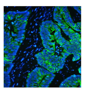
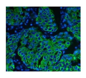
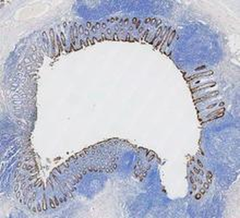
IF analysis of Cytokeratin 8/KRT8 using anti-Cytokeratin 8/KRT8 antibody. Cytokeratin 8/KRT8 was detected in a paraffin-embedded section of human appendix tissue. Heat mediated antigen retrieval was performed in EDTA buffer (pH8.0, epitope retrieval solution). The tissue section was blocked with 10% goat serum. The tissue section was then incubated with 5 µg/mL rabbit anti-Cytokeratin 8/KRT8 Antibody overnight at 4°C. DyLight 594 Conjugated AffiniPure Goat Anti-rabbit IgG (H+L) was used as secondary antibody at 1:100 dilution and incubated for 30 minutes at 37°C. The section was counterstained with DAPI. Visualize using a fluorescence microscope and filter sets appropriate for the label used.
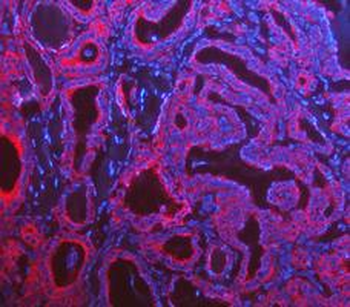
IF analysis of Cytokeratin 8/KRT8 using anti-Cytokeratin 8/KRT8 antibody. Cytokeratin 8/KRT8 was detected in a paraffin-embedded section of human ovarian cancer tissue. Heat mediated antigen retrieval was performed in EDTA buffer (pH8.0, epitope retrieval solution). The tissue section was blocked with 10% goat serum. The tissue section was then incubated with 5 µg/mL rabbit anti-Cytokeratin 8/KRT8 Antibody overnight at 4°C. DyLight 594 Conjugated AffiniPure Goat Anti-rabbit IgG (H+L) was used as secondary antibody at 1:100 dilution and incubated for 30 minutes at 37°C. The section was counterstained with DAPI. Visualize using a fluorescence microscope and filter sets appropriate for the label used.

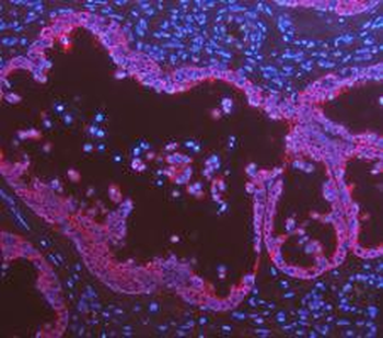
IF analysis of Cytokeratin 8/KRT8 using anti-Cytokeratin 8/KRT8 antibody. Cytokeratin 8/KRT8 was detected in a paraffin-embedded section of human placenta tissue. Heat mediated antigen retrieval was performed in EDTA buffer (pH8.0, epitope retrieval solution). The tissue section was blocked with 10% goat serum. The tissue section was then incubated with 5 µg/mL rabbit anti-Cytokeratin 8/KRT8 Antibody overnight at 4°C. DyLight 594 Conjugated AffiniPure Goat Anti-rabbit IgG (H+L) was used as secondary antibody at 1:100 dilution and incubated for 30 minutes at 37°C. The section was counterstained with DAPI. Visualize using a fluorescence microscope and filter sets appropriate for the label used.
IF analysis of Cytokeratin 8/KRT8 using anti-Cytokeratin 8/KRT8 antibody. Cytokeratin 8/KRT8 was detected in a paraffin-embedded section of human thyroid cancer tissue. Heat mediated antigen retrieval was performed in EDTA buffer (pH8.0, epitope retrieval solution). The tissue section was blocked with 10% goat serum. The tissue section was then incubated with 5 µg/mL rabbit anti-Cytokeratin 8/KRT8 Antibody overnight at 4°C. DyLight 594 Conjugated AffiniPure Goat Anti-rabbit IgG (H+L) was used as secondary antibody at 1:100 dilution and incubated for 30 minutes at 37°C. The section was counterstained with DAPI. Visualize using a fluorescence microscope and filter sets appropriate for the label used.
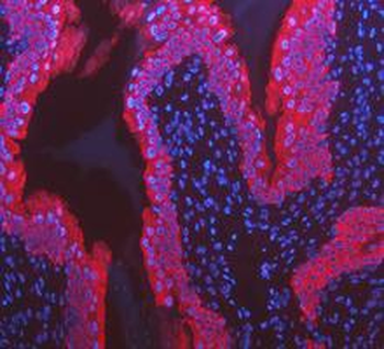
IF analysis of Cytokeratin 8/KRT8 using anti-Cytokeratin 8/KRT8 antibody. Cytokeratin 8/KRT8 was detected in a paraffin-embedded section of rat bladder cancer tissue. Heat mediated antigen retrieval was performed in EDTA buffer (pH8.0, epitope retrieval solution). The tissue section was blocked with 10% goat serum. The tissue section was then incubated with 5 µg/mL rabbit anti-Cytokeratin 8/KRT8 Antibody overnight at 4°C. DyLight 594 Conjugated AffiniPure Goat Anti-rabbit IgG (H+L) was used as secondary antibody at 1:100 dilution and incubated for 30 minutes at 37°C. The section was counterstained with DAPI. Visualize using a fluorescence microscope and filter sets appropriate for the label used.
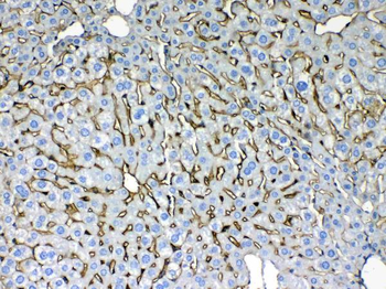
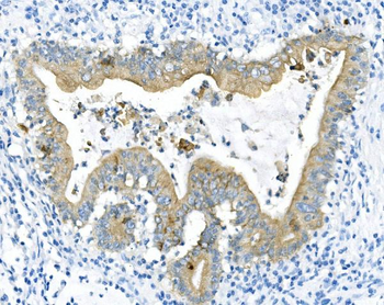
IHC analysis of Cytokeratin 8 using anti-Cytokeratin 8 antibody. Cytokeratin 8 was detected in paraffin-embedded section of mouse liver tissues. Heat mediated antigen retrieval was performed in citrate buffer (pH6, epitope retrieval solution) for 20 mins. The tissue section was blocked with 10% goat serum. The tissue section was then incubated with 1 µg/ml rabbit anti-Cytokeratin 8 Antibody overnight at 4°C. Biotinylated goat anti-rabbit IgG was used as secondary antibody and incubated for 30 minutes at 37°C. The tissue section was developed using Strepavidin-Biotin-Complex (SABC) with DAB as the chromogen.
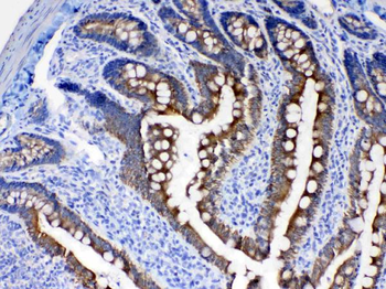
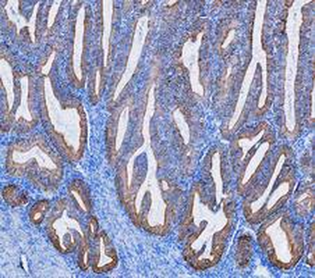
IHC analysis of Cytokeratin 8 using anti-Cytokeratin 8 antibody. Cytokeratin 8 was detected in paraffin-embedded section of rat intestine tissues. Heat mediated antigen retrieval was performed in citrate buffer (pH6, epitope retrieval solution) for 20 mins. The tissue section was blocked with 10% goat serum. The tissue section was then incubated with 1 µg/ml rabbit anti-Cytokeratin 8 Antibody overnight at 4°C. Biotinylated goat anti-rabbit IgG was used as secondary antibody and incubated for 30 minutes at 37°C. The tissue section was developed using Strepavidin-Biotin-Complex (SABC) with DAB as the chromogen.
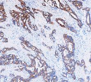
IHC analysis of Cytokeratin 8 using anti-Cytokeratin 8 antibody. Cytokeratin 8 was detected in paraffin-embedded section of human intestinal cancer tissues. Heat mediated antigen retrieval was performed in citrate buffer (pH6, epitope retrieval solution) for 20 mins. The tissue section was blocked with 10% goat serum. The tissue section was then incubated with 1 µg/ml rabbit anti-Cytokeratin 8 Antibody overnight at 4°C. Biotinylated goat anti-rabbit IgG was used as secondary antibody and incubated for 30 minutes at 37°C. The tissue section was developed using Strepavidin-Biotin-Complex (SABC) with DAB as the chromogen.
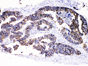
IHC analysis of Cytokeratin 8 using anti-Cytokeratin 8 antibody. Cytokeratin 8 was detected in paraffin-embedded section of human intestinal cancer tissues. Heat mediated antigen retrieval was performed in citrate buffer (pH6, epitope retrieval solution) for 20 mins. The tissue section was blocked with 10% goat serum. The tissue section was then incubated with 1 µg/ml rabbit anti-Cytokeratin 8 Antibody overnight at 4°C. Biotinylated goat anti-rabbit IgG was used as secondary antibody and incubated for 30 minutes at 37°C. The tissue section was developed using Strepavidin-Biotin-Complex (SABC) with DAB as the chromogen.
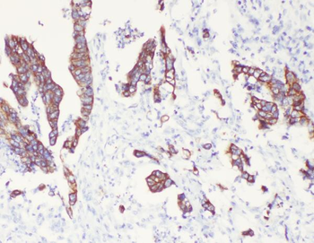
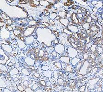
IHC analysis of Cytokeratin 8/KRT8 using anti-Cytokeratin 8/KRT8 antibody. Cytokeratin 8/KRT8 was detected in a paraffin-embedded section of human appendix cancer tissue. Heat mediated antigen retrieval was performed in EDTA buffer (pH8.0, epitope retrieval solution). The tissue section was blocked with 10% goat serum. The tissue section was then incubated with 1 µg/ml rabbit anti-Cytokeratin 8/KRT8 Antibody for 30 minutes at 37°C. HRP-AffiniPure Goat Anti-Rabbit IgG was used as secondary antibody and incubated for 30 minutes at 37°C. The tissue section was developed using HRP Conjugated Rabbit IgG Super Vision Assay Kit with DAB as the chromogen.
IHC analysis of Cytokeratin 8/KRT8 using anti-Cytokeratin 8/KRT8 antibody. Cytokeratin 8/KRT8 was detected in a paraffin-embedded section of human endometrial cancer tissue. Heat mediated antigen retrieval was performed in EDTA buffer (pH8.0, epitope retrieval solution). The tissue section was blocked with 10% goat serum. The tissue section was then incubated with 1 µg/ml rabbit anti-Cytokeratin 8/KRT8 Antibody for 30 minutes at 37°C. HRP-AffiniPure Goat Anti-Rabbit IgG was used as secondary antibody and incubated for 30 minutes at 37°C. The tissue section was developed using HRP Conjugated Rabbit IgG Super Vision Assay Kit with DAB as the chromogen.
IHC analysis of Cytokeratin 8/KRT8 using anti-Cytokeratin 8/KRT8 antibody. Cytokeratin 8/KRT8 was detected in a paraffin-embedded section of human ovarian cancer tissue. Heat mediated antigen retrieval was performed in EDTA buffer (pH8.0, epitope retrieval solution). The tissue section was blocked with 10% goat serum. The tissue section was then incubated with 1 µg/ml rabbit anti-Cytokeratin 8/KRT8 Antibody for 30 minutes at 37°C. HRP-AffiniPure Goat Anti-Rabbit IgG was used as secondary antibody and incubated for 30 minutes at 37°C. The tissue section was developed using HRP Conjugated Rabbit IgG Super Vision Assay Kit with DAB as the chromogen.
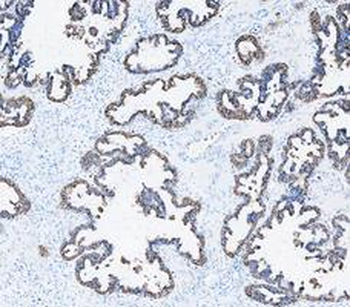
IHC analysis of Cytokeratin 8/KRT8 using anti-Cytokeratin 8/KRT8 antibody. Cytokeratin 8/KRT8 was detected in a paraffin-embedded section of human pancreas cancer tissue. Heat mediated antigen retrieval was performed in EDTA buffer (pH8.0, epitope retrieval solution). The tissue section was blocked with 10% goat serum. The tissue section was then incubated with 1 µg/ml rabbit anti-Cytokeratin 8/KRT8 Antibody for 30 minutes at 37°C. HRP-AffiniPure Goat Anti-Rabbit IgG was used as secondary antibody and incubated for 30 minutes at 37°C. The tissue section was developed using HRP Conjugated Rabbit IgG Super Vision Assay Kit with DAB as the chromogen.
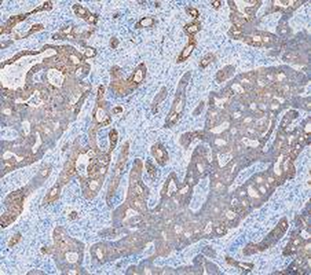
IHC analysis of Cytokeratin 8/KRT8 using anti-Cytokeratin 8/KRT8 antibody. Cytokeratin 8/KRT8 was detected in a paraffin-embedded section of human stomach cancer tissue. Heat mediated antigen retrieval was performed in EDTA buffer (pH8.0, epitope retrieval solution). The tissue section was blocked with 10% goat serum. The tissue section was then incubated with 1 µg/ml rabbit anti-Cytokeratin 8/KRT8 Antibody for 30 minutes at 37°C. HRP-AffiniPure Goat Anti-Rabbit IgG was used as secondary antibody and incubated for 30 minutes at 37°C. The tissue section was developed using HRP Conjugated Rabbit IgG Super Vision Assay Kit with DAB as the chromogen.
IHC analysis of Cytokeratin 8/KRT8 using anti-Cytokeratin 8/KRT8 antibody. Cytokeratin 8/KRT8 was detected in a paraffin-embedded section of human thyroid cancer tissue. Heat mediated antigen retrieval was performed in EDTA buffer (pH8.0, epitope retrieval solution). The tissue section was blocked with 10% goat serum. The tissue section was then incubated with 1 µg/ml rabbit anti-Cytokeratin 8/KRT8 Antibody for 30 minutes at 37°C. HRP-AffiniPure Goat Anti-Rabbit IgG was used as secondary antibody and incubated for 30 minutes at 37°C. The tissue section was developed using HRP Conjugated Rabbit IgG Super Vision Assay Kit with DAB as the chromogen.

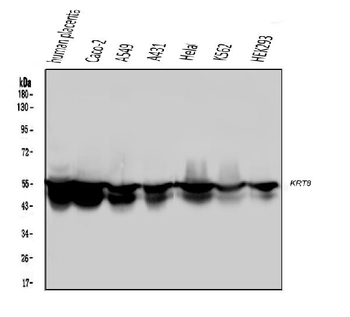
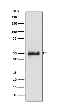
Western blot analysis of Cytokeratin 8 using anti-Cytokeratin 8 antibody. Electrophoresis was performed on a 5-20% SDS-PAGE gel at 70V (Stacking gel) / 90V (Resolving gel) for 2-3 hours. The sample well of each lane was loaded with 50 ug of sample under reducing conditions. Lane 1: rat liver tissue lysates, Lane 2: mouse ovary tissue lysates, Lane 3: mouse liver tissue lysates, Lane 4: MCF-7 whole cell lysates, Lane 5: A549 whole cell lysates, Lane 6: HELA whole Cell lysates. After Electrophoresis, proteins were transferred to a Nitrocellulose membrane at 150mA for 50-90 minutes. Blocked the membrane with 5% Non-fat Milk/ TBS for 1.5 hour at RT. The membrane was incubated with rabbit anti-Cytokeratin 8 antigen affinity purified polyclonal antibody at 0.5 µg/mL overnight at 4°C, then washed with TBS-0.1% Tween 3 times with 5 minutes each and probed with a goat anti-rabbit IgG-HRP secondary antibody at a dilution of 1:10000 for 1.5 hour at RT. The signal is developed using an Enhanced Chemiluminescent detection (ECL) kit with Tanon 5200 system. A specific band was detected for Cytokeratin 8 at approximately 54KD, 56KD. The expected band size for Cytokeratin 8 is at 54KD.
- Item 1 of 6
Anti-Cytokeratin 8 KRT8 Antibody (monoclonal, 3G9) [orb527059]
FC, ICC, IF, IHC, IHC-Fr, WB
Human, Mouse, Rat
Mouse
Monoclonal
Unconjugated
10 μg, 100 μg - Item 1 of 2
Anti-Cytokeratin 8/KRT8 Antibody [orb18145]
FC, IHC, WB
Human, Mouse, Rat
Rabbit
Polyclonal
Unconjugated
10 μg, 100 μg - Item 1 of 2
Anti-Cytokeratin 8 KRT8 Rabbit Monoclonal Antibody [orb547742]
FC, ICC, IF, IHC, IP, WB
Human, Mouse
Rabbit
Monoclonal
Unconjugated
30 μl, 100 μl 
Recombinant Cytokeratin 8 (KRT8) Antibody [orb751379]
IHC, WB
Human
Rabbit
Monoclonal
Unconjugated
100 μgRecombinant Cytokeratin 8 (KRT8) Antibody [orb751068]
IHC, WB
Human
Mouse
Monoclonal
Unconjugated
100 μg