You have no items in your shopping cart.
Cart summary
Item 1 of 3
Item 1 of 3
Anti-Collagen I/COL1A1 Antibody
Catalog Number: orb371673

| Catalog Number | orb371673 |
|---|---|
| Category | Antibodies |
| Description | Anti-Collagen I/COL1A1 Antibody |
| Species/Host | Rabbit |
| Clonality | Polyclonal |
| Tested applications | IHC, WB |
| Reactivity | Mouse, Rat |
| Isotype | Rabbit IgG |
| Immunogen | A synthetic peptide corresponding to a sequence at the C-terminus of mouse Collagen I, different from the related human sequence by three amino acids, and identical to the related rat sequence. |
| Concentration | Adding 0.2 ml of distilled water will yield a concentration of 500 μg/ml. |
| Form/Appearance | Lyophilized |
| Conjugation | Unconjugated |
| MW | 138-180 kDa |
| UniProt ID | P11087 |
| Storage | Maintain refrigerated at 2-8°C for up to 2 weeks. For long term storage store at -20°C in small aliquots to prevent freeze-thaw cycles. |
| Alternative names | Collagen alpha-1 (I) chain; Alpha-1 type I collage Read more... |
| Note | For research use only |
| Application notes | Immunohistochemistry (Paraffin-embedded Section), 0.5-1μg/ml, Mouse, Rat Western blot, 0.1-0.5μg/ml, Mouse. Add 0.2ml of distilled water will yield a concentration of 500ug/ml |
| Expiration Date | 12 months from date of receipt. |

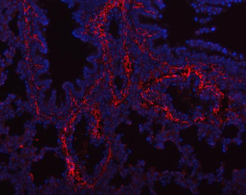
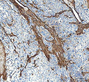
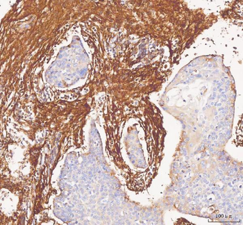
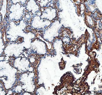
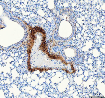
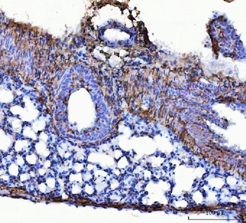
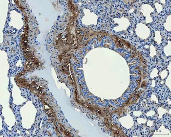
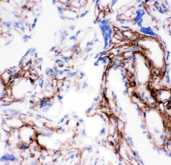
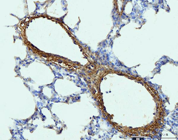
IHC analysis of COL1A1 using anti COL1A1 antibody. COL1A1 was detected in a paraffin-embedded section of mouse lung tissue. Heat mediated antigen retrieval was performed in EDTA buffer (pH8.0, epitope retrieval solution). The tissue section was blocked with 10% goat serum. The tissue section was then incubated with 2 µg/ml rabbit anti-COL1A1 Antibody overnight at 4°C. Peroxidase Conjugated Goat Anti-rabbit IgG was used as secondary antibody and incubated for 30 minutes at 37°C. The tissue section was developed using HRP Conjugated Rabbit IgG Super Vision Assay Kit with DAB as the chromogen.
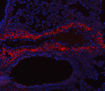
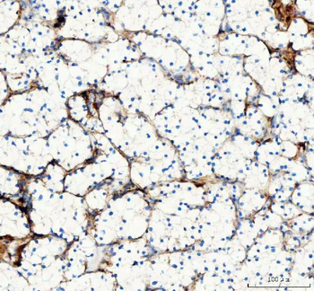
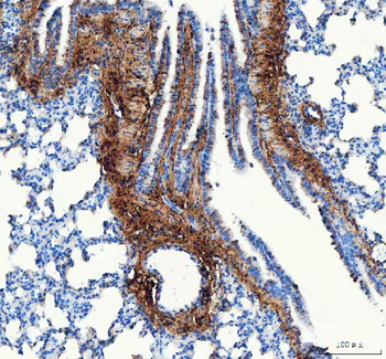
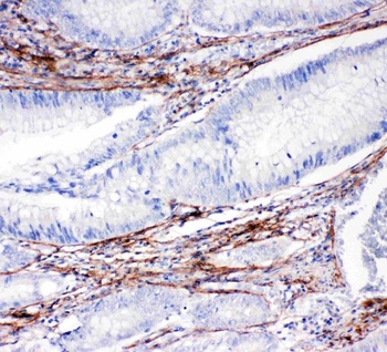
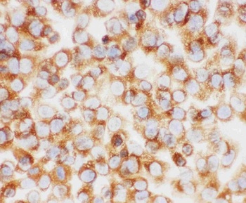
IHC analysis of COL1A1 using anti COL1A1 antibody. COL1A1 was detected in a paraffin-embedded section of rat lung tissue. Heat mediated antigen retrieval was performed in EDTA buffer (pH8.0, epitope retrieval solution). The tissue section was blocked with 10% goat serum. The tissue section was then incubated with 2 µg/ml rabbit anti-COL1A1 Antibody overnight at 4°C. Peroxidase Conjugated Goat Anti-rabbit IgG was used as secondary antibody and incubated for 30 minutes at 37°C. The tissue section was developed using HRP Conjugated Rabbit IgG Super Vision Assay Kit with DAB as the chromogen.
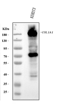
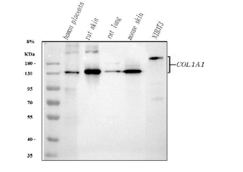
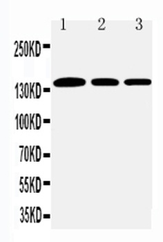
Western blot analysis of COL1A1 using anti-COL1A1 antibody. Electrophoresis was performed on a 5-20% SDS-PAGE gel at 70V (Stacking gel) / 90V (Resolving gel) for 2-3 hours. The sample well of each lane was loaded with 30 ug of sample under reducing conditions. Lane 1: mouse NIH/3T3 whole cell lysates. After electrophoresis, proteins were transferred to a nitrocellulose membrane at 150 mA for 50-90 minutes. Blocked the membrane with 5% non-fat milk/TBS for 1.5 hour at RT. The membrane was incubated with rabbit anti-COL1A1 antigen affinity purified polyclonal antibody at 0.5 µg/mL overnight at 4°C, then washed with TBS-0.1% Tween 3 times with 5 minutes each and probed with a goat anti-rabbit IgG-HRP secondary antibody at a dilution of 1:5000 for 1.5 hour at RT. The signal is developed using an Enhanced Chemiluminescent detection (ECL) kit with Tanon 5200 system. A specific band was detected for COL1A1 at approximately 138-180 kDa. The expected band size for COL1A1 is at 138 kDa.
- Item 1 of 11
Anti-Collagen I/COL1A1 Antibody [orb371672]
IF, IHC, WB
Human
Rabbit
Polyclonal
Unconjugated
10 μg, 100 μg - Item 1 of 8
Anti-Collagen I/COL1A1 Antibody [orb107158]
ICC, IF, IHC, WB
Human, Mouse, Rat
Rabbit
Polyclonal
Unconjugated
10 μg, 100 μg - Item 1 of 4
Anti-Collagen I/COL1A1 Antibody [orb654259]
IHC, WB
Human, Mouse
Rabbit
Polyclonal
Unconjugated
10 μg, 100 μg - Item 1 of 2
Anti-Collagen I COL1A1 Rabbit Monoclonal Antibody [orb547410]
IHC, WB
Human
Rabbit
Monoclonal
Unconjugated
30 μl, 100 μl 
Anti-Collagen I/COL1A1 Antibody [orb1882073]
ICC, IF, IHC, IHC-Fr, WB
Human, Mouse, Rat
Rabbit
Polyclonal
Unconjugated
100 μg