You have no items in your shopping cart.
Cart summary
Item 1 of 10
Item 1 of 10
Anti-CD31/Pecam1 Antibody Picoband
Catalog Number: orb402467

| Catalog Number | orb402467 |
|---|---|
| Category | Antibodies |
| Description | Anti-CD31/Pecam1 Antibody. Tested in ELISA, IHC, WB applications. This antibody reacts with Mouse. |
| Species/Host | Rabbit |
| Clonality | Polyclonal |
| Tested applications | ELISA, IHC, WB |
| Reactivity | Mouse |
| Isotype | Rabbit IgG |
| Immunogen | E. coli-derived mouse CD31 recombinant protein (Position: R58-Q273). |
| Concentration | Adding 0.2 ml of distilled water will yield a concentration of 500 μg/ml. |
| Form/Appearance | Lyophilized |
| Conjugation | Unconjugated |
| MW | 130 kDa |
| UniProt ID | Q08481 |
| Storage | Maintain refrigerated at 2-8°C for up to 2 weeks. For long term storage store at -20°C in small aliquots to prevent freeze-thaw cycles. |
| Alternative names | Platelet endothelial cell adhesion molecule; PECAM Read more... |
| Note | For research use only |
| Application notes | Western blot, 0.1-0.5μg/ml Immunohistochemistry, 2-5 μg/ml ELISA, 0.1-0.5μg/ml. Add 0.2ml of distilled water will yield a concentration of 500ug/ml |
| Expiration Date | 12 months from date of receipt. |

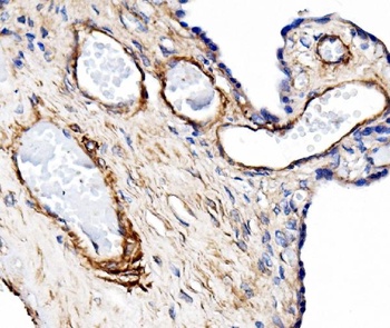
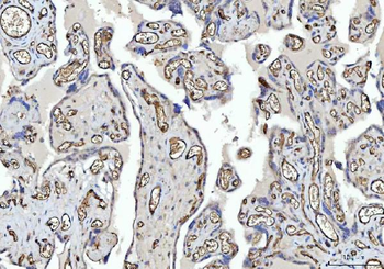
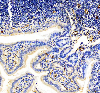
IHC analysis of CD31 using anti-CD31 antibody. CD31 was detected in a paraffin-embedded section of mouse colon tissue. Heat mediated antigen retrieval was performed in EDTA buffer (pH8.0, epitope retrieval solution). The tissue section was blocked with 10% goat serum. The tissue section was then incubated with 2 µg/ml rabbit anti-CD31 Antibody overnight at 4°C. Peroxidase Conjugated Goat Anti-rabbit IgG was used as secondary antibody and incubated for 30 minutes at 37°C. The tissue section was developed using HRP Conjugated Rabbit IgG Super Vision Assay Kit with DAB as the chromogen.
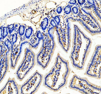
IHC analysis of CD31 using anti-CD31 antibody. CD31 was detected in a paraffin-embedded section of mouse colon tissue. Heat mediated antigen retrieval was performed in EDTA buffer (pH8.0, epitope retrieval solution). The tissue section was blocked with 10% goat serum. The tissue section was then incubated with 2 µg/ml rabbit anti-CD31 Antibody overnight at 4°C. Peroxidase Conjugated Goat Anti-rabbit IgG was used as secondary antibody and incubated for 30 minutes at 37°C. The tissue section was developed using HRP Conjugated Rabbit IgG Super Vision Assay Kit with DAB as the chromogen.
IHC analysis of CD31 using anti-CD31 antibody. CD31 was detected in a paraffin-embedded section of mouse colon tissue. Heat mediated antigen retrieval was performed in EDTA buffer (pH8.0, epitope retrieval solution). The tissue section was blocked with 10% goat serum. The tissue section was then incubated with 2 µg/ml rabbit anti-CD31 Antibody overnight at 4°C. Peroxidase Conjugated Goat Anti-rabbit IgG was used as secondary antibody and incubated for 30 minutes at 37°C. The tissue section was developed using HRP Conjugated Rabbit IgG Super Vision Assay Kit with DAB as the chromogen.
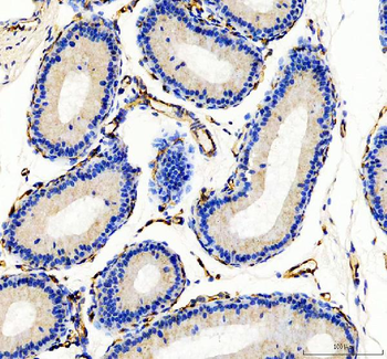
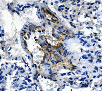
IHC analysis of CD31 using anti-CD31 antibody. CD31 was detected in a paraffin-embedded section of mouse epididymis tissue. Heat mediated antigen retrieval was performed in EDTA buffer (pH8.0, epitope retrieval solution). The tissue section was blocked with 10% goat serum. The tissue section was then incubated with 2 µg/ml rabbit anti-CD31 Antibody overnight at 4°C. Peroxidase Conjugated Goat Anti-rabbit IgG was used as secondary antibody and incubated for 30 minutes at 37°C. The tissue section was developed using HRP Conjugated Rabbit IgG Super Vision Assay Kit with DAB as the chromogen.
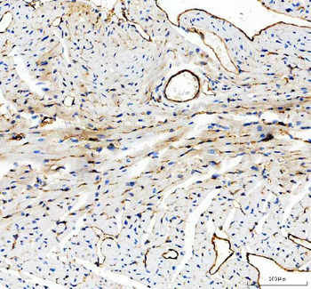
IHC analysis of CD31 using anti-CD31 antibody. CD31 was detected in a paraffin-embedded section of mouse heart tissue. Heat mediated antigen retrieval was performed in EDTA buffer (pH8.0, epitope retrieval solution). The tissue section was blocked with 10% goat serum. The tissue section was then incubated with 2 µg/ml rabbit anti-CD31 Antibody overnight at 4°C. Peroxidase Conjugated Goat Anti-rabbit IgG was used as secondary antibody and incubated for 30 minutes at 37°C. The tissue section was developed using HRP Conjugated Rabbit IgG Super Vision Assay Kit with DAB as the chromogen.
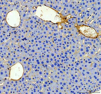
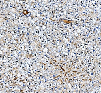
IHC analysis of CD31 using anti-CD31 antibody. CD31 was detected in a paraffin-embedded section of mouse liver tissue. Heat mediated antigen retrieval was performed in EDTA buffer (pH8.0, epitope retrieval solution). The tissue section was blocked with 10% goat serum. The tissue section was then incubated with 2 µg/ml rabbit anti-CD31 Antibody overnight at 4°C. Peroxidase Conjugated Goat Anti-rabbit IgG was used as secondary antibody and incubated for 30 minutes at 37°C. The tissue section was developed using HRP Conjugated Rabbit IgG Super Vision Assay Kit with DAB as the chromogen.
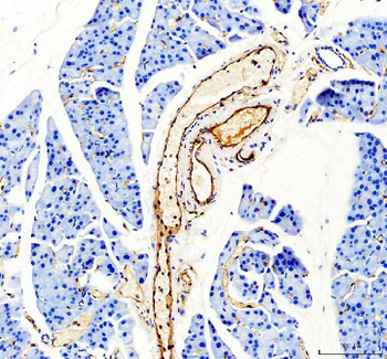
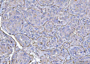
IHC analysis of CD31 using anti-CD31 antibody. CD31 was detected in a paraffin-embedded section of mouse pancrease tissue. Heat mediated antigen retrieval was performed in EDTA buffer (pH8.0, epitope retrieval solution). The tissue section was blocked with 10% goat serum. The tissue section was then incubated with 2 µg/ml rabbit anti-CD31 Antibody overnight at 4°C. Peroxidase Conjugated Goat Anti-rabbit IgG was used as secondary antibody and incubated for 30 minutes at 37°C. The tissue section was developed using HRP Conjugated Rabbit IgG Super Vision Assay Kit with DAB as the chromogen.
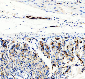
IHC analysis of CD31 using anti-CD31 antibody. CD31 was detected in a paraffin-embedded section of mouse stomach tissue. Heat mediated antigen retrieval was performed in EDTA buffer (pH8.0, epitope retrieval solution). The tissue section was blocked with 10% goat serum. The tissue section was then incubated with 2 µg/ml rabbit anti-CD31 Antibody overnight at 4°C. Peroxidase Conjugated Goat Anti-rabbit IgG was used as secondary antibody and incubated for 30 minutes at 37°C. The tissue section was developed using HRP Conjugated Rabbit IgG Super Vision Assay Kit with DAB as the chromogen.
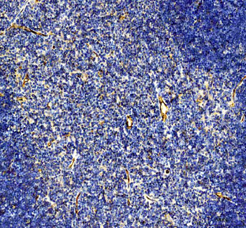
IHC analysis of CD31 using anti-CD31 antibody. CD31 was detected in a paraffin-embedded section of mouse thymus tissue. Heat mediated antigen retrieval was performed in EDTA buffer (pH8.0, epitope retrieval solution). The tissue section was blocked with 10% goat serum. The tissue section was then incubated with 2 µg/ml rabbit anti-CD31 Antibody overnight at 4°C. Peroxidase Conjugated Goat Anti-rabbit IgG was used as secondary antibody and incubated for 30 minutes at 37°C. The tissue section was developed using HRP Conjugated Rabbit IgG Super Vision Assay Kit with DAB as the chromogen.
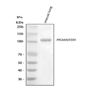
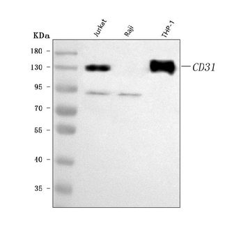
Western blot analysis of CD31 using anti-CD31 antibody. Electrophoresis was performed on a 5-20% SDS-PAGE gel at 70V (Stacking gel) / 90V (Resolving gel) for 2-3 hours. The sample well of each lane was loaded with 30 ug of sample under reducing conditions. Lane 1: mouse lung tissue lysates. After electrophoresis, proteins were transferred to a nitrocellulose membrane at 150 mA for 50-90 minutes. Blocked the membrane with 5% non-fat milk/TBS for 1.5 hour at RT. The membrane was incubated with rabbit anti-CD31 antigen affinity purified polyclonal antibody at 0.5 µg/mL overnight at 4°C, then washed with TBS-0.1% Tween 3 times with 5 minutes each and probed with a goat anti-rabbit IgG-HRP secondary antibody at a dilution of 1:5000 for 1.5 hour at RT. The signal is developed using an Enhanced Chemiluminescent detection (ECL) kit with Tanon 5200 system. A specific band was detected for CD31 at approximately 120-130 kDa. The expected band size for CD31 is at 83 kDa.
- Item 1 of 5
Anti-CD31/PECAM1 Antibody Picoband (monoclonal, 2D4) [orb654278]
IHC, IHC-Fr, WB
Human
Mouse
Monoclonal
Unconjugated
10 μg, 100 μg - Item 1 of 4
Anti-CD31/Pecam1 Antibody Picoband [orb623816]
ELISA, IHC, WB
Human, Mouse, Rat
Rabbit
Polyclonal
Unconjugated
100 μg, 10 μg - Item 1 of 4
Anti-CD31/PECAM1 Antibody Picoband [orb196264]
FC, IHC, WB
Human
Rabbit
Polyclonal
Unconjugated
10 μg, 100 μg - Item 1 of 3
Anti-CD31/PECAM1 Antibody Picoband [orb137862]
FC, IF, IHC, WB
Human
Rabbit
Polyclonal
Unconjugated
10 μg, 100 μg - Item 1 of 2
Anti-CD31/Pecam1 Antibody Picoband [orb623815]
ELISA, FC, WB
Mouse, Rat
Rabbit
Polyclonal
Unconjugated
100 μg, 10 μg