You have no items in your shopping cart.
Cart summary
Item 1 of 4
Item 1 of 4
Anti-Cd27 Antibody
Catalog Number: orb1098031

| Catalog Number | orb1098031 |
|---|---|
| Category | Antibodies |
| Description | Anti-Cd27 Antibody. Tested in ELISA, Flow Cytometry, IHC applications. This antibody reacts with Mouse, Rat. |
| Species/Host | Rabbit |
| Clonality | Polyclonal |
| Tested applications | ELISA, FC, IHC |
| Reactivity | Mouse, Rat |
| Isotype | Rabbit IgG |
| Immunogen | E.coli-derived mouse Cd27 recombinant protein (Position: P24-F187). |
| Antibody Type | Primary Antibody |
| Concentration | Adding 0.2 ml of distilled water will yield a concentration of 500 μg/ml. |
| Form/Appearance | Lyophilized |
| Conjugation | Unconjugated |
| MW | 45 kDa |
| UniProt ID | P41272 |
| Storage | Maintain refrigerated at 2-8°C for up to 2 weeks. For long term storage store at -20°C in small aliquots to prevent freeze-thaw cycles. |
| Alternative names | Angiopoietin-related protein 4; Angiopoietin-like Read more... |
| Note | For research use only |
| Application notes | Immunohistochemistry(Paraffin-embedded Section), 2-5 μg/ml, Mouse, Rat Flow Cytometry (Fixed), 1-3 μg/1x106 cells, Mouse ELISA, 0.1-0.5 μg/ml, -. Adding 0.2 ml of distilled water will yield a concentration of 500 μg/ml |
| Expiration Date | 12 months from date of receipt. |

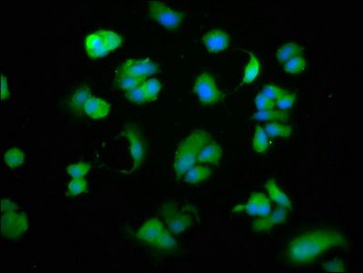
Flow Cytometry analysis of mouse PBMC cells using anti-Cd27 antibody. Overlay histogram showing mouse PBMC cells (Blue line). The cells were fixed with 4% paraformaldehyde and blocked with 10% normal goat serum. And then incubated with rabbit anti-Cd27 Antibody (1 µg/1x10^6 cells) for 30 min at 20°C. DyLight®488 conjugated goat anti-rabbit IgG (5-10 µg/1x10^6 cells) was used as secondary antibody for 30 minutes at 20°C. Isotype control antibody (Green line) was rabbit IgG (1 µg/1x10^6) used under the same conditions. Unlabelled sample without incubation with primary antibody and secondary antibody (Red line) was used as a blank control.
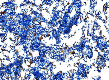
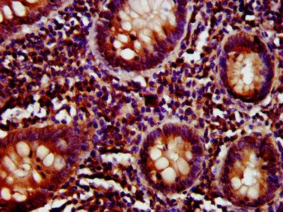
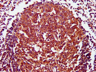
IHC analysis of Cd27 using anti-Cd27 antibody. Cd27 was detected in a paraffin-embedded section of mouse lymphaden tissue. Heat mediated antigen retrieval was performed in EDTA buffer (pH8.0, epitope retrieval solution). The tissue section was blocked with 10% goat serum. The tissue section was then incubated with 2 µg/ml rabbit anti-Cd27 Antibody overnight at 4°C. Peroxidase Conjugated Goat Anti-rabbit IgG was used as secondary antibody and incubated for 30 minutes at 37°C. The tissue section was developed using HRP Conjugated Rabbit IgG Super Vision Assay Kit with DAB as the chromogen.
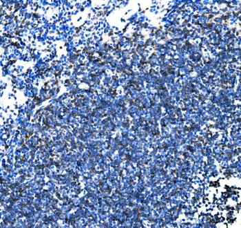
IHC analysis of Cd27 using anti-Cd27 antibody. Cd27 was detected in a paraffin-embedded section of rat lymphaden tissue. Heat mediated antigen retrieval was performed in EDTA buffer (pH8.0, epitope retrieval solution). The tissue section was blocked with 10% goat serum. The tissue section was then incubated with 2 µg/ml rabbit anti-Cd27 Antibody overnight at 4°C. Peroxidase Conjugated Goat Anti-rabbit IgG was used as secondary antibody and incubated for 30 minutes at 37°C. The tissue section was developed using HRP Conjugated Rabbit IgG Super Vision Assay Kit with DAB as the chromogen.
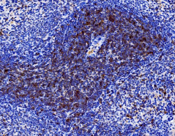
IHC analysis of Cd27 using anti-Cd27 antibody. Cd27 was detected in a paraffin-embedded section of rat spleen tissue. Heat mediated antigen retrieval was performed in EDTA buffer (pH8.0, epitope retrieval solution). The tissue section was blocked with 10% goat serum. The tissue section was then incubated with 2 µg/ml rabbit anti-Cd27 Antibody overnight at 4°C. Peroxidase Conjugated Goat Anti-rabbit IgG was used as secondary antibody and incubated for 30 minutes at 37°C. The tissue section was developed using HRP Conjugated Rabbit IgG Super Vision Assay Kit with DAB as the chromogen.
- Item 1 of 6
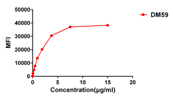
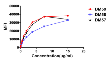
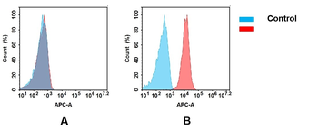
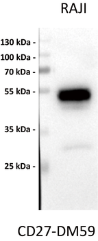
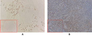
Anti-CD27 antibody(DM59), Rabbit mAb [orb757382]
ELISA, FC, IHC, WB
Human
Rabbit
Monoclonal
Unconjugated
50 μg, 100 μg, 500 μg, 10 μg - Item 1 of 4
- Item 1 of 4
Anti-CD27 antibody(DM57); Rabbit mAb [orb757380]
ELISA, FC
Human
Rabbit
Monoclonal
Unconjugated
10 μg, 50 μg, 100 μg, 500 μg - Item 1 of 4
Anti-CD27 antibody(DM58); Rabbit mAb [orb757381]
ELISA, FC
Human
Rabbit
Monoclonal
Unconjugated
50 μg, 100 μg, 500 μg, 10 μg - Item 1 of 2


![Anti-CD27 [LG.3A10]](/images//pub/media/catalog/product/NewWebsite/35/orb613904_1.png)
![Anti-CD27 [LG.3A10]](/images/pub/media/catalog/product/NewWebsite/35/orb613904_2.png)
