You have no items in your shopping cart.
Cart summary
Item 1 of 4
Item 1 of 4
Anti-beta-Tubulin Purified
Catalog Number: orb44535

| Catalog Number | orb44535 |
|---|---|
| Category | Antibodies |
| Description | Mouse Monoclonal to beta tubulin. |
| Clonality | Monoclonal |
| Clone Number | TU-06 |
| Tested applications | ICC, IHC-P, WB |
| Reactivity | Fish, Gallus, Human, Mouse, Other, Paramecium, Plant, Porcine, Rat |
| Isotype | Mouse IgM |
| Immunogen | Beta-subunits of porcine brain tubulin. |
| Antibody Type | Primary Antibody |
| Concentration | 1 mg/ml |
| Purity | Purified by sequential steps of physicochemical fractionation (differential precipitation and solid-phase chromatography methods). |
| Conjugation | Unconjugated |
| Target | beta-Tubulin |
| Entrez | 81027 |
| UniProt ID | Q9H4B7 |
| RRID | AB_10990495 |
| Storage | Maintain refrigerated at 2-8°C for up to 2 weeks. For long term storage store at -20°C in small aliquots to prevent freeze-thaw cycles. |
| Buffer/Preservatives | Tris buffered saline (TBS), pH 8.0, 15 mM sodium azide |
| Alternative names | Anti-beta-tubulin antibody Read more... |
| Note | For research use only |
| Application notes | Immunocytochemistry: Recommended dilution: 2 μg/ml, fixed and permeabilized cells; positive control: 3T3 mouse embryonal fibroblast cell line. Immunohistochemistry (paraffin sections): Recommended dilution: 5 μg/ml, positive tissue: heart. Western blotting: Recommended dilution: 1-2 μg/ml. |
| Expiration Date | 12 months from date of receipt. |

Immunocytochemistry staining of 3T3 mouse embryonal fibroblast cell line using anti-beta-tubulin (TU-06) (detection by Goat anti-mouse IgM Cy®5). Nucleus is stained with DAPI (blue).

Immunohistochemistry staining of human heart (paraffin sections) using anti-beta tubulin (TU-06).

Western blotting analysis of human beta-tubulin using mouse monoclonal antibody TU-06 on lysates (50 mM TRIS-Cl pH 6.8, 4M UREA, 4% SDS) of various cell lines under non-reducing and reducing conditions. Nitrocellulose membrane was probed with 2 µg/ml of mouse anti-beta-tubulin monoclonal antibody followed by IRDye800-conjugated anti-mouse secondary antibody. A specific band was detected for beta-tubulin at approximately 54 kDa.

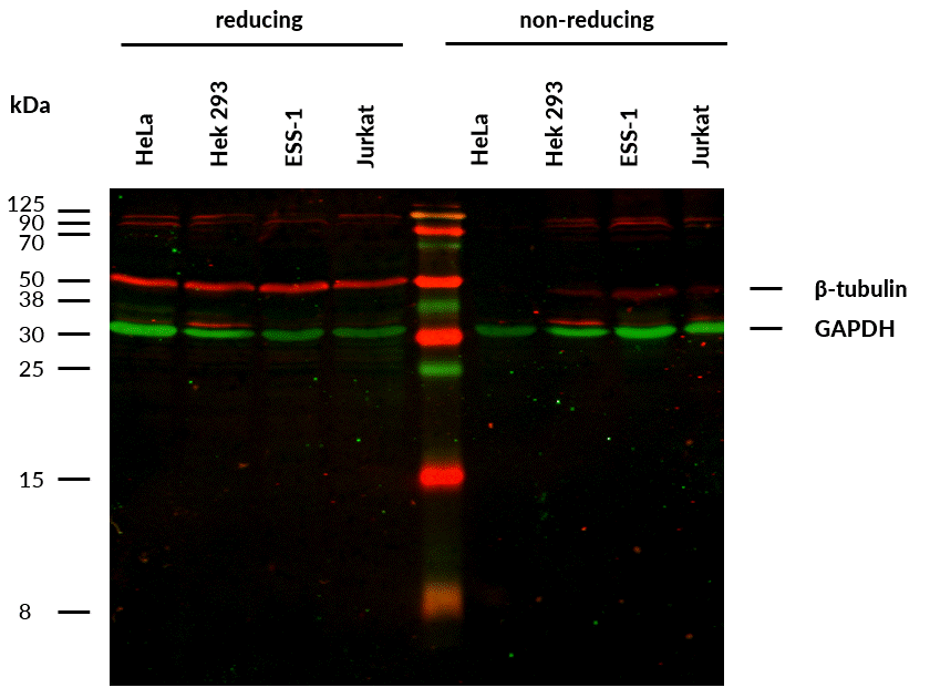
Anti-beta-Tubulin Purified (TU-06) works in WB application under reducing conditions on RIPA cell extracts. Western blotting analysis was performed on whole cell extracts (RIPA lysis buffer) of HeLa, HEK 293, ESS-1 and Jurkat cell lines mixed and heated (100°C, 5 min) with reducing (2-mercaptoethanol) or non-reducing SDS-loading buffer. Samples were resolved using 12% Tris-glycine SDS gel electrophoresis. Nitrocellulose membrane blot was probed simultaneously with mouse IgM monoclonal antibody TU-06 (1 µg/ml) and mouse IgG1 anti-GAPDH monoclonal antibody FF26A (1 µg/ml) used as the loading control. Subclass-specific secondary antibodies IRDye 680RD Goat-anti-Mouse IgM (red) and IRDye 800CW Goat-anti-Mouse IgG (green) were used for multiplex fluorescent Western blot detection. Alpha-tubulin was detected at ~50 kDa in all tested cell lines under reducing, but not under non- reducing conditions. Using RIPA lysis buffer in combination with non-reducing conditions is not suitable for Anti-beta-Tubulin Purified (TU-06).
- Item 1 of 2
Anti-alpha/beta-Tubulin dimer Purified [orb44537]
FC, ICC, WB
Human, Mouse, Porcine, Primate, Rat
Monoclonal
Unconjugated
0.1 mg - Item 1 of 1
Anti-alpha/beta-Tubulin dimer Purified [orb44536]
ICC, WB
Human, Mouse, Porcine
Monoclonal
Unconjugated
0.1 mg - Item 1 of 1
Anti-beta-Tubulin Purified [orb44542]
ICC, IHC-Fr, WB
Human, Mouse, Plant, Porcine
Monoclonal
Unconjugated
0.1 mg - Item 1 of 1
Anti-beta-Tubulin Purified [orb44541]
FC, ICC, WB
Human, Mouse, Plant, Porcine, Rat
Monoclonal
Unconjugated
0.1 mg 
Anti-Beta Tubulin III (Tuj-1) Antibody [orb1821784]
WB
Human, Mouse, Rat
Rabbit
Polyclonal
Unconjugated
100 μl