You have no items in your shopping cart.
Cart summary
Item 1 of 6
Item 1 of 6
Anti-alpha-Tubulin Purified
Catalog Number: orb44543

| Catalog Number | orb44543 |
|---|---|
| Category | Antibodies |
| Description | Mouse Monoclonal to alpha tubulin. |
| Clonality | Monoclonal |
| Clone Number | TU-16 |
| Tested applications | ELISA, ICC, IHC-P, IP, WB |
| Reactivity | Canine, Human, Mouse, Plant, Porcine, Rat |
| Isotype | Mouse IgM |
| Immunogen | Porcine brain microtubule protein MTP-1. |
| Concentration | 1 mg/ml |
| Purity | Purified by sequential steps of physicochemical fractionation (differential precipitation and solid-phase chromatography methods). |
| Conjugation | Unconjugated |
| Target | alpha-Tubulin |
| Entrez | 7277 |
| UniProt ID | Q71U36 |
| RRID | AB_11012737 |
| Storage | Maintain refrigerated at 2-8°C for up to 2 weeks. For long term storage store at -20°C in small aliquots to prevent freeze-thaw cycles. |
| Buffer/Preservatives | Tris buffered saline (TBS), pH 8.0, 15 mM sodium azide |
| Alternative names | Anti-alpha-tubulin antibody Read more... |
| Note | For research use only |
| Application notes | Western blotting: Recommended dilution: 1-2 μg/ml; positive control: HPB-ALL human peripheral blood leukemia cell line, reducing conditions. Immunohistochemistry (paraffin sections): Recommended dilution: 10 μg/ml. Immunoprecipitation: Reducing conditions. |
| Expiration Date | 12 months from date of receipt. |

Immunoprecipitation of alpha-tubulin from HeLa and RAJI cell lysate by antibody TU-16 and its detection by antibody TU-01. IgM heavy chain (76-92 kDa) and IgM light chain (25-30 kDa) indicated. Mr of alpha tubulin is around 50 kDa. L = lysate; IPr = immunoprecipitate (reducing conditions).

Immunocytochemistry staining of alpha-tubulin in Hep-2 cells using mouse monoclonal antibody TU-16 (diluted 1:400), detected with GAM IgG-Alexa Fluor®488 (diluted 1:200; green).

Immunohistochemistry staining of human heart (paraffin sections) using anti-alpha tubulin (TU-16).

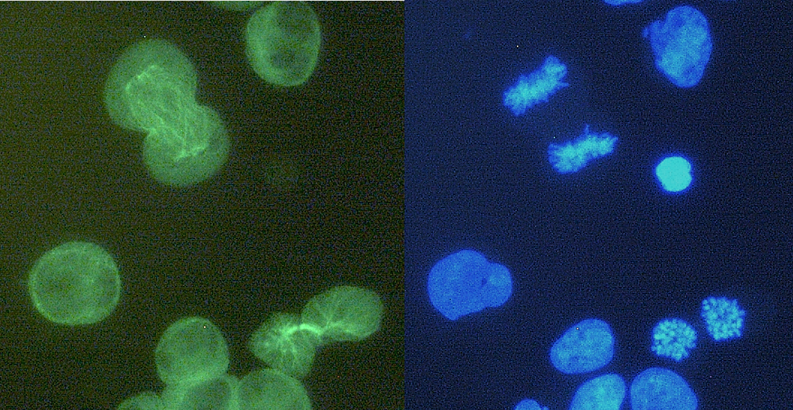
Immunohistochemistry staining (paraffin sections) of alpha-tubulin in human stomach using mouse monoclonal antibody TU-16 (diluted 1:400), detected with GAM IgG-Alexa Fluor®488 (diluted 1:200; green).

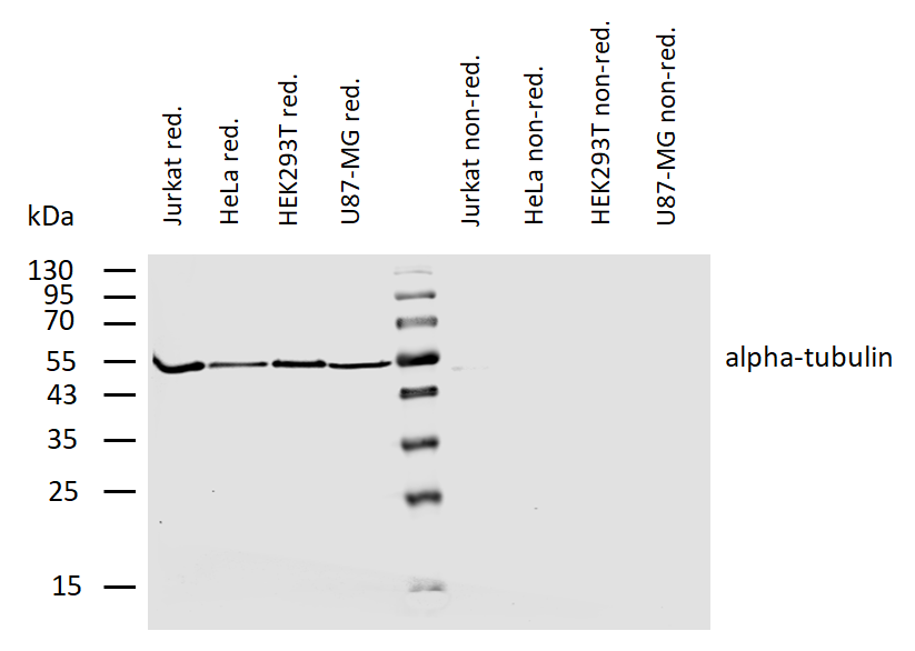
Western blotting analysis of human alpha-tubulin using mouse monoclonal antibody TU-16 on lysates of various cell lines and porcine brain under reducing and non-reducing conditions. Nitrocellulose membrane was probed with 2 µg/ml of mouse anti-alpha-tubulin monoclonal antibody followed by IRDye800-conjugated anti-mouse secondary antibody. A specific band was detected for alpha-tubulin at approximately 54 kDa, nonspecific minor bands above 100 kDa do not interfere with specific signal.

Anti-alpha-Tubulin Purified (TU-16) works in WB application under reducing conditions. Western blotting analysis was performed on whole cell extracts (RIPA lysis buffer) of HeLa, HEK 293, ESS-1 and Jurkat cell lines mixed and heated (100°C, 5 min) with reducing (2-mercaptoethanol) or non-reducing SDS-loading buffer. Samples were resolved using 12% Tris-glycine SDS gel electrophoresis. Nitrocellulose membrane blot was probed simultaneously with mouse IgM monoclonal antibody TU-16 (1 µg/ml) and mouse IgG1 anti-GAPDH monoclonal antibody FF26A (1 µg/ml) used as the loading control. Subclass-specific secondary antibodies IRDye 680RD Goat-anti-Mouse IgM (red) and IRDye 800CW Goat-anti-Mouse IgG (green) were used for multiplex fluorescent Western blot detection. Alpha-tubulin was detected at ~50 kDa in all tested cell lines.
- Item 1 of 9
Anti-alpha-Tubulin Purified [orb44529]
FC, ICC, IHC-P, IP, WB
Aves, Human, Invertebrate, Mouse, Paramecium, Plant, Porcine, Yeast
Monoclonal
Unconjugated
0.1 mg - Item 1 of 2
- Item 1 of 2
Anti-alpha-Tubulin Purified [orb738510]
ELISA, FC, WB
Aves, Human, Mammal, Mouse, Rat, Yeast
Monoclonal
Unconjugated
0.1 mg - Item 1 of 3
Rabbit anti-alpha-Tubulin Antibody, Affinity Purified [orb1520420]
IP, WB
Human, Mouse
Rabbit
Polyclonal
Unconjugated
10 μg - Item 1 of 3
Rabbit anti-alpha-Tubulin Antibody, Affinity Purified [orb1520421]
IP, WB
Human, Mouse
Rabbit
Polyclonal
Unconjugated
100 μg