You have no items in your shopping cart.
Cart summary
Item 1 of 5
Item 1 of 5
Angpt1 antibody
Catalog Number: orb750479

| Catalog Number | orb750479 |
|---|---|
| Category | Antibodies |
| Description | Angpt1 antibody |
| Species/Host | Rabbit |
| Clonality | Polyclonal |
| Tested applications | ELISA, IF, IHC, WB |
| Reactivity | Human, Mouse |
| Isotype | Antiserum |
| Immunogen | This whole rabbit serum was prepared by repeated immunizations with a synthetic peptide corresponding to a N-terminus region near aa 15-45 of mouse angiopoietin 1 protein conjugated to KLH using maleimide. A residue of cysteine was added to the amino terminal end to facilitate coupling. |
| Concentration | 80 mg/ml |
| Dilution range | ELISA: 1:5,000 - 1:25,000, IHC: 1:200-1:800, WB: 1:500-1:2,000 |
| Form/Appearance | Liquid (sterile filtered) |
| Purity | Anti-Angiopoietin 1 is directed against mouse angiopoietin-1 and shows no reactivity with mouse angiopoietin-2. This reagent cross-reacts with human angiopoietin 1. Partially cross reactivity is noted with human angiopoietin 2. This product was prepared from monospecific antiserum by a delipidation and defibrination. |
| Conjugation | Unconjugated |
| UniProt ID | O08538 |
| NCBI | 46048213 |
| Storage | Store vial at -20° C or below prior to opening. This vial contains a relatively low volume of reagent (25 µL). To minimize loss of volume dilute 1:10 by adding 225 µL of the buffer stated above directly to the vial. Recap, mix thoroughly and briefly centrifuge to collect the volume at the bottom of the vial. Use this intermediate dilution when calculating final dilutions as recommended below. Store the vial at -20°C or below after dilution. Avoid cycles of freezing and thawing. |
| Buffer/Preservatives | 0.01% (w/v) Sodium Azide |
| Alternative names | rabbit anti-Angiopoietin 1 Antibody, rabbit anti-A Read more... |
| Note | For research use only |
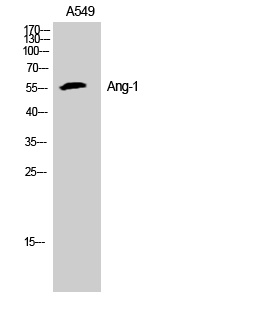
| Application notes | Anti-Antiopoietin-1 Antiserum has been tested in ELISA, western blot, immunohistochemistry, and immunofluorescence and is suitable for other antibody based assays. A 1:500 dilution is recommended for western blotting. The reaction of this antiserum directly with cell supernatants may result in high background due to reactivity of components in the serum. This can be alleviated by first immunoprecipitating the antibody:antigen complex and then detecting the antigen. This method results in a very clean and strong signal. Both Ang-1 and Ang-2 proteins have predicted molecular weights of approximately 57 kDa and appear on western blots close to their predicted molecular weights. In some instances additional bands may be seen at approximately 75 kDa which represent highly glycosylated forms of the protein that migrate at a higher apparent molecular weight. |
| Expiration Date | 12 months from date of receipt. |

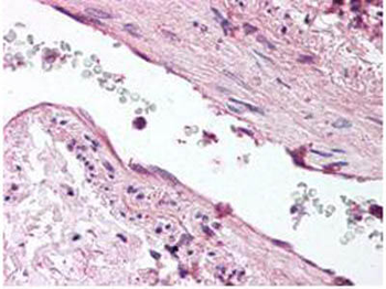
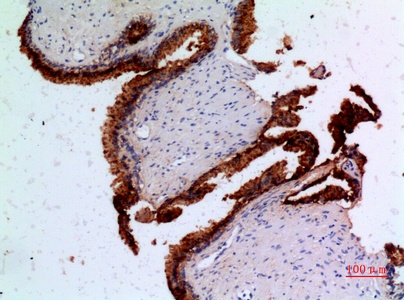
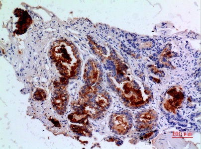
Biorbyt's anti-ANG1 antibody was diluted 1:500 to detect ANG1 in human lung tissue. Tissue was formalin fixed and paraffin embedded. No pre-treatment of sample was required. The image shows the localization of antibody as the precipitated red signal, with a hematoxylin purple nuclear counter stain.

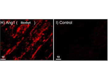
Immunofluorescence and confocal imaging of Rabbit Anti-Angiopoietin 1 Antibody. (H) Perfusion-fixed mouse Left Ventricle. Anti-Angiopoietin 1 conjugated to Alexa Fluor 568 (red), which recognizes only monomers, immunostains CMs and ECs in vivo. I) Rabbit sera (negative control). Scale bars = 10 μm.
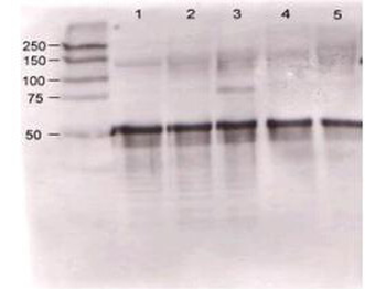
Rabbit anti-Ang-1 was used at a 1:500 dilution to detect mouse Ang-1 by western blot against supernatants of mouse angiopoietin-expressing endothelial cells. Lane 1 - wt endothelial cells. Lane 2 - mouse Ang-1 (clone 1-8) expressing cells. Lane 3 - mouse Ang-1 (clone 1-15) expressing cells. Lane 4 - mouse Ang-2 (clone 2-9) expressing cells. Approximately 20 µg of each lysate was used for 10% SDS-PAGE. Immunoprecipitation preceded the reaction with primary antibody at room temperature for 1 h. After subsequent washing, a 1:5000 dilution of HRP conjugated Gt-a-Rabbit IgG (orb347654) preceded color development.

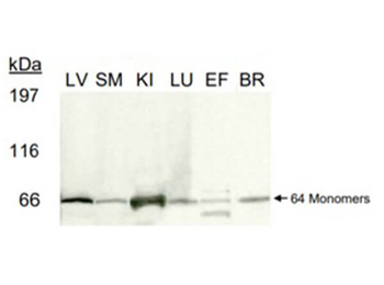
Western blot of Rabbit Anti-Angiopoietin 1 Antibody. Tested on 100 µg of PNGase-F-treated and reduced lysates: Mouse Left Ventricle, Skeletal Muscle, Kidney, Lung, Epididymal Fat Tissue, and Brain on a 3-8% gel. Anti-Angiopoietin 1 incubated for 1 hr, anti-Rabbit IgG HRP secondary 1:1500 incubated for 1hr.
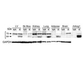
Western blot of Rb Anti-Angiopoietin 1 Antibody. 100 µg of protein lysates treated with PNGase F and evaluated under reduced conditions from Human Left Ventricle, skeletal muscle, kidney, lung, epididymal fat tissue, and brain tissue. Anti-Angiopoietin1 incubated and reprobed with Anti-GAPDH antibodies.
- Item 1 of 5
ANGPT1 antibody [orb22485]
ELISA, FC, IF, WB
Bovine, Canine, Human, Mouse, Porcine, Rat
Goat
Polyclonal
Unconjugated
100 μg - Item 1 of 5
- Item 1 of 4
- Item 1 of 3
- Item 1 of 3
ANGPT1 Antibody [orb1563485]
ELISA, IHC-P, WB
Human, Rat
Rabbit
Polyclonal
Unconjugated
100 μl, 50 μl, 20 μl


Submit a review
Filter by Rating

- 5 stars

- 4 stars
- 3 stars
- 2 stars
- 1 stars
