You have no items in your shopping cart.
Cart summary
Item 1 of 6
Item 1 of 6
ALPL Antibody (Center)
Catalog Number: orb1935523

| Catalog Number | orb1935523 |
|---|---|
| Category | Antibodies |
| Description | Purified Rabbit Polyclonal Antibody (Pab) |
| Species/Host | Rabbit |
| Clonality | Polyclonal |
| Clone Number | RB14192 |
| Tested applications | FC, IF, IHC-P, WB |
| Reactivity | Human, Mouse |
| Isotype | Rabbit IgG |
| Antibody Type | Primary Antibody |
| Dilution range | IF: 1:10~50, WB: 1:2000, WB: 1:2000, IHC-P-Leica: 1:500, IHC-P-Leica: 1:500, FC: 1:10~50 |
| Form/Appearance | Purified polyclonal antibody supplied in PBS with 0.09% (W/V) sodium azide. This antibody is purified through a protein A column, followed by peptide affinity purification. |
| Conjugation | Unconjugated |
| MW | 57305 Da |
| Target | This ALPL antibody is generated from rabbits immunized with a KLH conjugated synthetic peptide between 217-246 amino acids from the Central region of human ALPL. |
| UniProt ID | P05186 |
| NCBI | NP_001170991.1, NP_001120973.2, NP_000469.3 |
| Storage | Maintain refrigerated at 2-8°C for up to 2 weeks. For long term storage store at -20°C in small aliquots to prevent freeze-thaw cycles |
| Alternative names | Alkaline phosphatase, tissue-nonspecific isozyme, Read more... |
| Note | For research use only |
| Expiration Date | 12 months from date of receipt. |

Confocal immunofluorescent analysis of ALPL Antibody (Center) with MCF-7 cell followed by Alexa Fluor 488-conjugated goat anti-rabbit lgG (green). DAPI was used to stain the cell nuclear (blue).
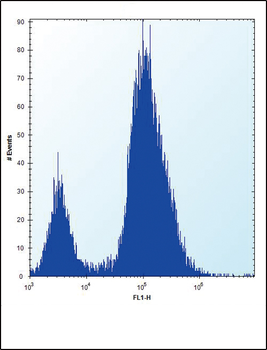
ALPL Antibody (Center) flow cytometric analysis of 293 cells (right histogram) compared to a negative control cell (left histogram). FITC-conjugated goat-anti-rabbit secondary antibodies were used for the analysis.
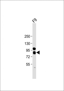
Anti-ALPL Antibody (Center) at 1:2000 dilution + F9 whole cell lysate. Lysates/proteins at 20 µg per lane. Secondary Goat Anti-Rabbit IgG, (H+L), Peroxidase conjugated at 1/10000 dilution. Predicted band size: 57 kDa. Blocking/Dilution buffer: 5% NFDM/TBST.
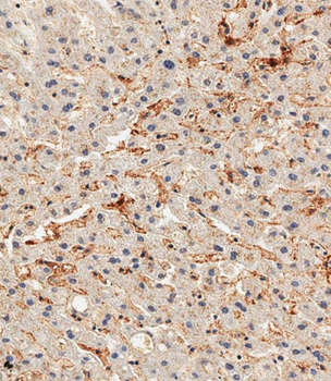
Immunohistochemical analysis of paraffin-embedded human liver tissue was performed on the Leica BOND RXm. Samples were incubated with primary antibody (1/500) for 1 hours at room temperature. A undiluted biotinylated CRF Anti-Polyvalent HRP Polymer antibody was used as the secondary antibody.
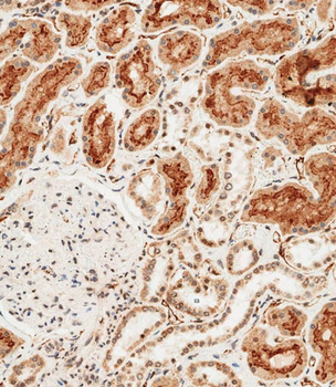
Immunohistochemical analysis of paraffin-embedded human kidney tissue was performed on the Leica BOND RXm. Samples were incubated with primary antibody (1/500) for 1 hours at room temperature. A undiluted biotinylated CRF Anti-Polyvalent HRP Polymer antibody was used as the secondary antibody.
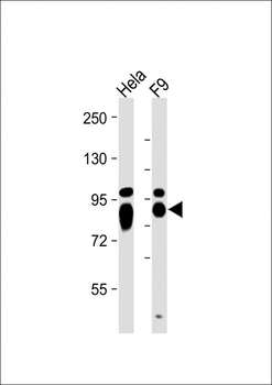
All lanes: Anti-ALPL Antibody (Center) at 1:2000 dilution. Lane 1: Hela whole cell lysate. Lane 2: F9 whole cell lysate. Lysates/proteins at 20 µg per lane. Secondary Goat Anti-Rabbit IgG, (H+L), Peroxidase conjugated at 1/10000 dilution. Predicted band size: 57 kDa. Blocking/Dilution buffer: 5% NFDM/TBST.
