You have no items in your shopping cart.
Cart summary
Item 1 of 5
Item 1 of 5
Alpha Synuclein Antibody (pSer129): Biotin
Catalog Number: orb414136

| Catalog Number | orb414136 |
|---|---|
| Category | Antibodies |
| Description | Rabbit polyclonal antibody against Alpha Synuclein pSer129 conjugated to Biotin |
| Species/Host | Rabbit |
| Clonality | Polyclonal |
| Tested applications | ELISA, ICC, IF, IHC, WB |
| Reactivity | Human, Mouse, Rat |
| Immunogen | Synthetic peptide of Human Alpha Synuclein pSer129 (124-134 aa), conjugated to Keyhole Limpet Haemocyanin (KLH). |
| Concentration | 0.5mg/mL |
| Dilution range | WB (1:1000); ICC/IF (1:500) |
| Conjugation | Biotin |
| MW | 100, 75, 45, 15 kDa |
| Target | Alpha Synuclein pSer129 |
| Entrez | 6622 |
| UniProt ID | P37840 |
| NCBI | NP_000336.1 |
| Storage | Conjugated antibodies should be stored according to the product label |
| Buffer/Preservatives | 136.36mM Ethanolamine, and 9.55mM Sodium Bicarbonate in 95.45% PBS |
| Alternative names | Phospho anti-alpha Synuclein (S129) antibody, Alph Read more... |
| Note | For research use only |
| Application notes | A 1:1000 dilution of SPC-742 was sufficient for detection of Alpha Synuclein pSer129 in 15 µg of human brain cell lysates by ECL immunoblot analysis using goat anti-rabbit IgG:HRP as the secondary antibody. |
| Expiration Date | 12 months from date of receipt. |

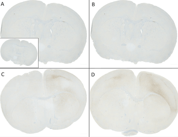
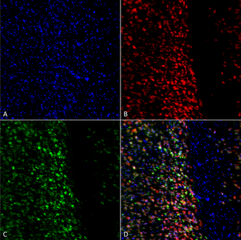
Immunohistochemistry analysis using Rabbit Anti-Alpha Synuclein (pSer129) Polyclonal Antibody. Tissue: Free floating brain sections. Species: Mouse. Fixation: PFA. Primary Antibody: Rabbit Anti-Alpha Synuclein (pSer129) Polyclonal Antibody at 1:500 for overnight at 4°C with gentle agitation. Counterstain: Hoechst. Magnification: 63X. A) Right hemisphere (striatum) injected with alpha synuclein AAV vector. B) Control. Alpha synuclein streaks are visible at injection site, but not control, 4 months after injection.
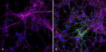
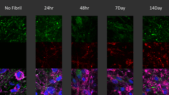
Immunocytochemistry/Immunofluorescence analysis using Rabbit Anti-Alpha Synuclein (pSer129) Polyclonal Antibody. Species: Mouse. Primary Antibody: Rabbit Anti-Alpha Synuclein (pSer129) Polyclonal Antibody. Phospho serine 129 antibody was used to detect phosphorylated alpha synuclein in primary mouse hippocampal neurons treated with 100 nM sonicated mouse alpha synuclein PFFs (A). Phosphorylated alpha synuclein was visible in perinucleus and neurites compared to untreated control (B).
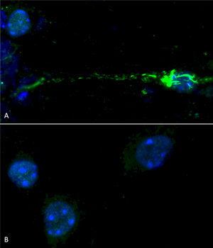
Immunocytochemistry/Immunofluorescence analysis using Rabbit Anti-Alpha Synuclein pSer129 Polyclonal Antibody. Tissue: Primary hippocampal neurons treated with active Alpha Synuclein Protein Aggregate at 4 μg/ml to induce fibrils. Species: Rat. Fixation: 4% paraformaldehyde. Primary Antibody: Rabbit Anti-Alpha Synuclein pSer129 Polyclonal Antibody at 1:500 for 24 hours at 4°C. Secondary Antibody: Goat Anti-Rabbit Alexa Fluor 488 at 1:700 for 1 hour at RT. Counterstain: Guinea Pig Anti-NeuN (red) neuronal marker (Donkey Anti-Guinea Pig Alexa Fluor 647 1:700); Hoechst (blue) nuclear stain at 1:6000, 1:3000 for 60 min at RT, 5 min at RT. Magnification: 20X.
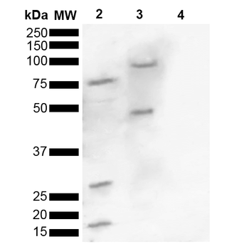
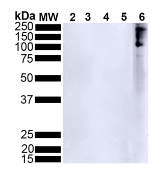
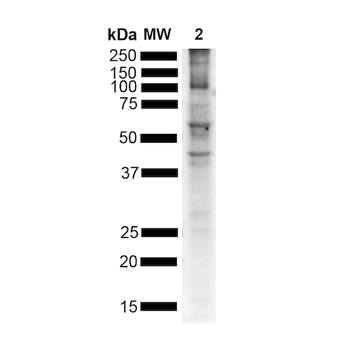
Western blot analysis of Human, Mouse brain lysate showing detection of ~16 kDa Alpha Synuclein pSer129 protein using Rabbit Anti-Alpha Synuclein pSer129 Polyclonal Antibody. Lane 1: Molecular Weight Ladder (MW). Lane 2: Human brain lysate. Lane 3: Mouse brain lysate. Lane 4: Human Alpha Synuclein Monomer (0.5 μg). Load: 15 μg. Block: 5% Skim Milk in 1X TBST. Primary Antibody: Rabbit Anti-Alpha Synuclein pSer129 Polyclonal Antibody at 1:1000 for 2 hours at RT. Secondary Antibody: Goat Anti-Rabbit HRP:IgG at 1:3000 for 1 hour at RT. Color Development: ECL solution for 5 min at RT. Predicted/Observed Size: ~16 kDa. Other Band (s): 100, 75, 45, 30, 16 kDa.
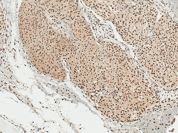
Immunohistochemistry analysis using Rabbit Anti-Alpha Synuclein pSer129 Polyclonal Antibody. Tissue: Brain. Species: Human. Fixation: Formalin Fixed Paraffin-Embedded. Primary Antibody: Rabbit Anti-Alpha Synuclein pSer129 Polyclonal Antibody at 1:50 for 30 min at RT. Counterstain: Hematoxylin. Magnification: 10X. HRP-DAB Detection.
- Item 1 of 7
Alpha Synuclein Antibody (pSer129): Biotin [orb612725]
ELISA, ICC, IF, IHC, WB
Human, Mouse
Rabbit
Monoclonal
Biotin
100 μg