You have no items in your shopping cart.
Cart summary
Item 1 of 5
Item 1 of 5
Alpha Synuclein Antibody: Biotin
Catalog Number: orb413308

| Catalog Number | orb413308 |
|---|---|
| Category | Antibodies |
| Description | Mouse monoclonal antibody against alpha Synuclein conjugated to Biotin |
| Species/Host | Mouse |
| Clonality | Monoclonal |
| Clone Number | 3C11 |
| Tested applications | ELISA, ICC, IF, IHC, WB |
| Reactivity | Human, Mouse, Rat |
| Isotype | IgG1 |
| Immunogen | Human alpha synuclein monomer |
| Concentration | 1 mg/ml |
| Dilution range | WB (1:1000); DB (1:1000); ICC/IF (1:100); ELISA (1:1000) |
| Conjugation | Biotin |
| MW | 30 kDa |
| Target | Alpha Synuclein |
| Entrez | 6622 |
| UniProt ID | P37840 |
| NCBI | NP_000336.1 |
| Storage | Conjugated antibodies should be stored according to the product label |
| Buffer/Preservatives | 136.36mM Ethanolamine, and 9.55mM Sodium Bicarbonate in 95.45% PBS |
| Alternative names | Alpha Synuclein antibody, Non-A beta component of Read more... |
| Note | For research use only |
| Application notes | A 1:1000 dilution of SMC-530 was sufficient for detection of Alpha Synuclein in 15 µg of human brain cell lysate by ECL immunoblot analysis using goat anti-mouse IgG:HRP as the secondary antibody. |
| Expiration Date | 12 months from date of receipt. |

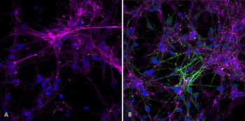
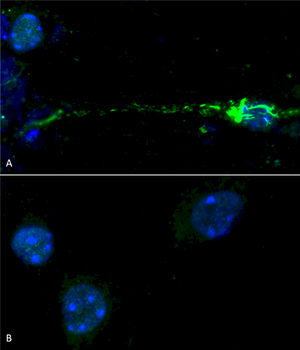
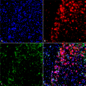
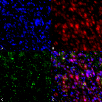
Immunocytochemistry/Immunofluorescence analysis using Mouse Anti-Alpha Synuclein Monoclonal Antibody, Clone 3C11. Tissue: Neuroblastoma cell line (SK-N-BE). Species: Human. Fixation: 4% Formaldehyde for 15 min at RT. Primary Antibody: Mouse Anti-Alpha Synuclein Monoclonal Antibody at 1:100 for 60 min at RT. Secondary Antibody: Goat Anti-Mouse ATTO 488 at 1:200 for 60 min at RT. Counterstain: Phalloidin Texas Red F-Actin stain; DAPI (blue) nuclear stain at 1:1000, 1:5000 for 60 min at RT, 5 min at RT. Localization: Cytoplasm: weak; Nucleus: Med. Magnification: 60X. (A) DAPI (blue) nuclear stain. (B) Phalloidin Texas Red F-Actin stain. (C) Alpha Synuclein Antibody. (D) Composite.
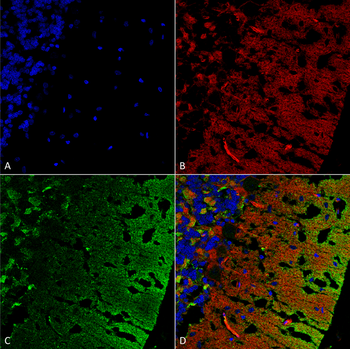
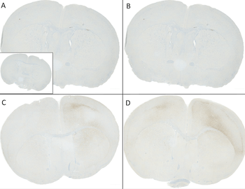
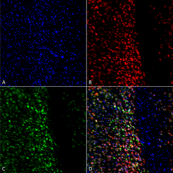
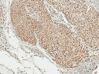
Immunohistochemistry analysis using Mouse Anti-Alpha Synuclein Monoclonal Antibody, Clone 3C11. Tissue: cerebellum. Species: Rat. Fixation: Formalin fixed, paraffin embedded. Primary Antibody: Mouse Anti-Alpha Synuclein Monoclonal Antibody at 1:25 for 1 hour at RT. Secondary Antibody: Goat Anti-Mouse IgG: Alexa Fluor 488. Counterstain: Actin-binding Phalloidin-Alexa Fluor 633; DAPI (blue) nuclear stain. Magnification: 63X. (A) DAPI (blue) nuclear stain. (B) Phalloidin Alexa Fluor 633 F-Actin stain. (C) Alpha Synuclein Antibody (D) Composite.
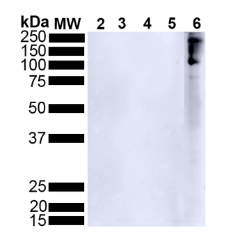
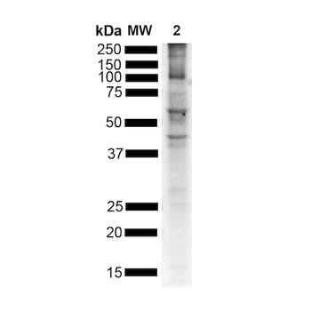
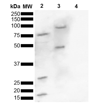
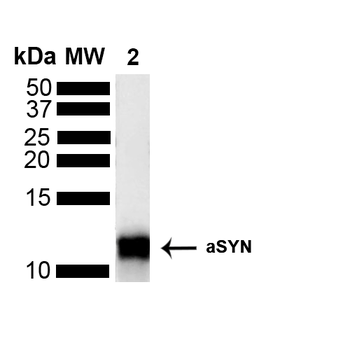
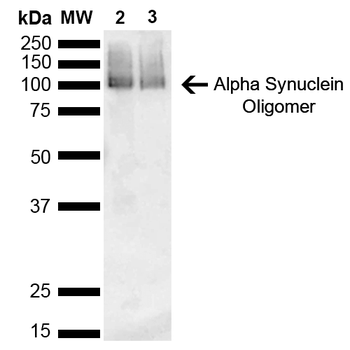
Western Blot analysis of Human Brain showing detection of 14 kDa Alpha Synuclein protein using Mouse Anti-Alpha Synuclein Monoclonal Antibody, Clone 3C11. Lane 1: Molecular Weight Ladder (MW). Lane 2: Parkinson brain cell lystate. Lane 3: Human brain cell lysate. Load: 15 μg. Block: 5% Skim Milk in 1X TBST. Primary Antibody: Mouse Anti-Alpha Synuclein Monoclonal Antibody at 1:1000 for 2 hours at RT. Secondary Antibody: Goat Anti-Mouse HRP:IgG at 1:3000 for 1 hour at RT. Color Development: ECL solution (Super Signal West Pico) for 5 min in RT. Predicted/Observed Size: 14 kDa. Other Band (s): 100 kDa (oligomer).
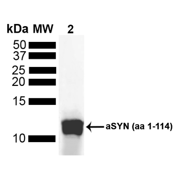
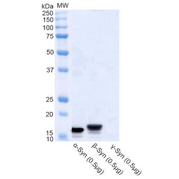
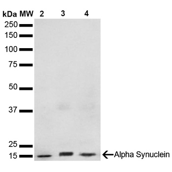
Western Blot analysis of Human Truncated Alpha Synuclein Protein showing detection of Alpha Synuclein protein using Mouse Anti-Alpha Synuclein Monoclonal Antibody, Clone 3C11. Lane 1: MW Ladder. Lane 2: hASYN aa 1-114 (5 uL). Load: 5 uL. Block: 5% Skim Milk Powder in TBST. Primary Antibody: Mouse Anti-Alpha Synuclein Monoclonal Antibody at 1:1000 for 2 hours at RT with shaking. Secondary Antibody: Goat anti-mouse IgG:HRP at 1:4000 for 1 hour at RT with shaking. Color Development: Chemiluminescent for HRP (Moss) for 5 min in RT.
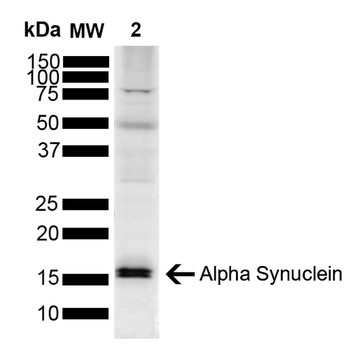
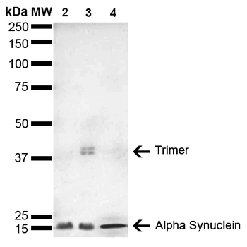
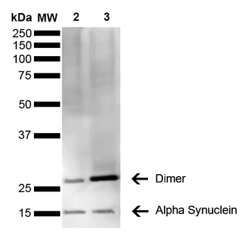
Western Blot analysis of Mouse, Rat Brain showing detection of 14 kDa Alpha Synuclein protein using Mouse Anti-Alpha Synuclein Monoclonal Antibody, Clone 3C11. Lane 1: Molecular Weight Ladder (MW). Lane 2: Mouse brain cell lysate. Lane 3: Rat brain cell lysate. Load: 15 μg. Block: 5% Skim Milk in 1X TBST. Primary Antibody: Mouse Anti-Alpha Synuclein Monoclonal Antibody at 1:1000 for 2 hours at RT. Secondary Antibody: Goat Anti-Mouse HRP:IgG at 1:3000 for 1 hour at RT. Color Development: ECL solution (Super Signal West Pico) for 5 min in RT. Predicted/Observed Size: 14 kDa. Other Band (s): ~30 kDa (dimer).
- Item 1 of 7
Alpha Synuclein Antibody (pSer129): Biotin [orb612725]
ELISA, ICC, IF, IHC, WB
Human, Mouse
Rabbit
Monoclonal
Biotin
100 μg - Item 1 of 5
Alpha Synuclein Antibody (pSer129): Biotin [orb414136]
ELISA, ICC, IF, IHC, WB
Human, Mouse, Rat
Rabbit
Polyclonal
Biotin
100 μl - Item 1 of 4
Alpha Synuclein Antibody: Biotin [orb536155]
ELISA, ICC, IF, IHC, WB
Human, Mouse, Rat
Rabbit
Polyclonal
Biotin
100 μg - Item 1 of 3
Alpha Synuclein Antibody: Biotin [orb413344]
ELISA, ICC, IF, IHC, WB
Human, Mouse, Rat
Mouse
Monoclonal
Biotin
100 μg - Item 1 of 3
Alpha Synuclein Antibody: Biotin [orb413362]
ELISA, ICC, IF, IHC, WB
Human, Mouse, Rat
Mouse
Monoclonal
Biotin
100 μg