You have no items in your shopping cart.
Cart summary
Item 1 of 4
Item 1 of 4
Aldolase antibody
Catalog Number: orb344330

| Catalog Number | orb344330 |
|---|---|
| Category | Antibodies |
| Description | Aldolase antibody |
| Species/Host | Goat |
| Clonality | Polyclonal |
| Tested applications | ELISA, IP, WB |
| Reactivity | Human, Rabbit |
| Isotype | IgG |
| Immunogen | Aldolase [Rabbit Muscle] |
| Concentration | 1 mg/ml |
| Dilution range | ELISA: 1:7,000, IP: 1:100, WB: 1:1,000 - 1:5,000 |
| Form/Appearance | Lyophilized |
| Purity | Anti-ALDOLASE was prepared from monospecific antiserum by a delipidation, salt fractionation and ion exchange chromatography. Assay by immunoelectrophoresis resulted in a single precipitin arc against anti-Goat Serum, purified and partially purified Aldolase [Rabbit Muscle]. |
| Conjugation | Unconjugated |
| UniProt ID | P00883 |
| NCBI | NP_001075707.1 |
| Storage | Store vial at 4° C prior to restoration. For extended storage aliquot contents and freeze at -20° C or below. Avoid cycles of freezing and thawing. Centrifuge product if not completely clear after standing at room temperature. This product is stable for several weeks at 4° C as an undiluted liquid. Dilute only prior to immediate use. |
| Buffer/Preservatives | 0.01% (w/v) Sodium Azide |
| Alternative names | goat anti-Aldolase Antibody, Fructose-bisphosphate Read more... |
| Note | For research use only |
| Application notes | Anti-Aldolase Antibody has been tested by ELISA, immunoprecipitation, and western blot. This product is assayed against 1.0 µg of Aldolase [Rabbit Muscle] in a standard ELISA using Peroxidase conjugated Affinity Purified anti-Goat IgG [H&L] (Rabbit) code #605-4302 and (ABTS (2,2’-azino-bis-[3-ethylbenthiazoline-6-sulfonic acid]) code # ABTS-100 as a substrate for 30 minutes at room temperature. A working dilution of 1:10,000 to 1:40,000 is suggested for this product. Use approximately 5 ul of antibody to immunoprecipitate 50 ul of protein lysate. |
| Expiration Date | 12 months from date of receipt. |
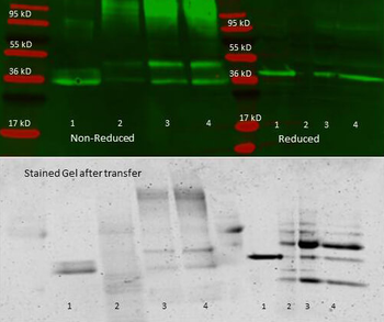
Anti aldolase antibody – Immunoprecipitation and Western Blot. 300 µL aliquots of whole anti-aldolase antiserum (orb750415) were used to precipitate varying amounts of purified aldolase and precipitates with controls were compared by SDS-PAGE and Western blot. Samples shown in the image are: 1. Purified aldolase 2. 300 µL antiserum with no antigen (negative control) 3. 300 µL antiserum with ~100 µL aldolase (2.5 mg/mL) 4. 300 µL antiserum with ~200 µL aldolase (2.5 mg/mL) For the precipitation, 300 ul of antiserum and an equal volume of aldolase antigen in PBS was incubated ~24 hrs at 4°C, centrifuged for 6 minutes at 13K RPM, washed once with PBS, centrifuged and dissolved in 60 ul 0.1 N NaOH. 90 ul of PBS was added, the sample was divided in 2 portions, and an equal volume of reducing (+ 4% BME) or non-reducing 2X sample buffer was added. The reduced samples were boiled for five minutes, and all samples were run at 140 V for ~45 minutes on a 4-20% tris/glycine gradient gel. Gel was stained, destained and imaged using standard protocols. Precipitation of aldolase was confirmed by comparison of increasing amounts of antigen with the control protein by SDS PAGE and observation of a 40-45 kD MW band corresponding to Aldolase. Additional higher and lower molecular weight bands correspond to serum proteins.
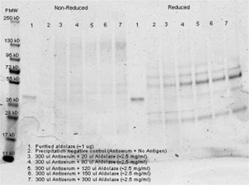
Anti aldolase antibody– Immunoprecipitation- Immunoprecipitation was performed with 300 ul of anti aldolase antiserum and an equal volume of varied amounts (diluted from a stock solution of ~2.5 mg/mL) of purified aldolase in PBS. Antibody/Antigen mixture was incubated ~24 hrs at 4°C, centrifuged for 6 minutes at 13K RPM, washed once with PBS, centrifuged and dissolved in 60 ul 0.1 N NaOH. 90 ul of PBS was added, the sample was divided in 2 portions, and an equal volume of reducing (+ 4% BME) or non-reducing 2X sample buffer was added. The reduced samples were boiled for five minutes, and all samples were run at 140 V for ~45 minutes on a 4-20% tris/glycine gradient gel. Gel was stained, destained and imaged using standard protocols. Precipitation of aldolase was confirmed by comparison of increasing amounts of antigen with the control protein by SDS PAGE and observation of a 40-45 kD MW band corresponding to Aldolase. Additional higher and lower molecular weight bands correspond to serum proteins.
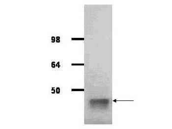
IgG purified antibody to rabbit muscle aldolase was used at a 1:1000 dilution to detect human aldolase by Western blot. A whole cell lysate prepared from human derived A293 cells was loaded on a 4-12% tris glycine gradient gel for SDS-PAGE. The gel was transferred to nitro-cellulose using standard techniques. Antibody reaction with the membrane occurred overnight at 4°C in TTBS supplemented with 2% non-fat dry milk. Color was allowed to develop using SuperSignal West Pico Chemiluminescent Substrate (PIERCE). Other detection methods will yield similar results. This antibody clearly detects a band at ~41 kDa consistent with human aldolase.
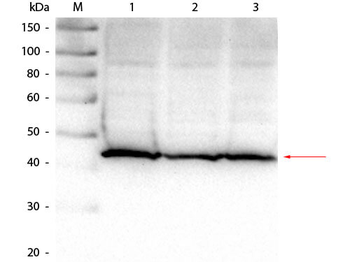
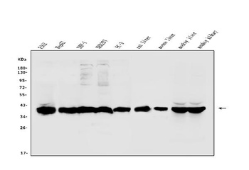
Western Blot of Goat anti-Aldolase Antibody. Lane 1: Hela lysate (p/n orb348668). Lane 2: HEK293 lysate (p/n orb348669). Lane 3: Jurkat lysate (p/n orb348674). Load: 25 µg per lane. Primary antibody: Goat anti-Aldolase Antibody at 1:1000 overnight at 4°C. Secondary antibody: HRP Dk-a-Gt IgG secondary antibody (p/n orb347031) at 1:40000 for 30 min at RT. Block: (p/n orb348637) for 30 min at RT. Predicted/Observed size: 39 kDa, 41 kDa for Aldolase.
- Item 1 of 9
Aldolase/ALDOA Antibody [orb570364]
ELISA, FC, ICC, IF, IHC, WB
Human, Mouse, Rat
Rabbit
Polyclonal
Unconjugated
10 μg, 100 μg - Item 1 of 7
Aldolase/ALDOB Antibody [orb570365]
ELISA, FC, ICC, IF, IHC, WB
Human, Monkey, Mouse, Rat
Rabbit
Polyclonal
Unconjugated
10 μg, 100 μg - Item 1 of 6
Aldolase/ALDOA Antibody (monoclonal, 6H8) [orb692226]
FC, ICC, IF, IHC, WB
Human
Mouse
Monoclonal
Unconjugated
10 μg, 100 μg - Item 1 of 4
- Item 1 of 4
Steroid 17-alpha-hydroxylase/17, 20 lyase antibody [orb240144]
ELISA, IF, IHC, WB
Human
Rabbit
Polyclonal
Unconjugated
100 μg, 50 μg


Submit a review
Filter by Rating

- 5 stars

- 4 stars
- 3 stars
- 2 stars
- 1 stars
