You have no items in your shopping cart.
Cart summary
Item 1 of 4
Item 1 of 4
ALDH1A1 Antibody
Catalog Number: orb1268147

| Catalog Number | orb1268147 |
|---|---|
| Category | Antibodies |
| Description | ALDH1A1 Antibody |
| Species/Host | Rabbit |
| Clonality | Polyclonal |
| Tested applications | FC, IF, IHC-P, WB |
| Reactivity | Human |
| Isotype | Rabbit Ig |
| Immunogen | This ALDH1A1 antibody is generated from rabbits immunized with human ALDH1A1 recombinant protein. |
| Concentration | batch dependent |
| Dilution range | For WB starting dilution is: 1:1000For IF starting dilution is: 1:100For IHC-P starting dilution is: 1:50~100For FACS starting dilution is: 1:10~50 |
| Form/Appearance | Liquid |
| Conjugation | Unconjugated |
| MW | 55 kDa |
| Target | ALDH1A1 |
| UniProt ID | P00352 |
| NCBI | P00352 |
| Storage | Store at 4°C for three months and -20°C, stable for up to one year. As with all antibodies care should be taken to avoid repeated freeze thaw cycles. Antibodies should not be exposed to prolonged high temperatures. |
| Buffer/Preservatives | Supplied in PBS with 0.09% (W/V) sodium azide. |
| Alternative names | Retinal dehydrogenase 1, RALDH 1, RalDH1, ALDH-E1, Read more... |
| Note | For research use only |
| Application notes | For WB starting dilution is: 1:1000For IF starting dilution is: 1:100For IHC-P starting dilution is: 1:50~100For FACS starting dilution is: 1:10~50 |
| Expiration Date | 12 months from date of receipt. |
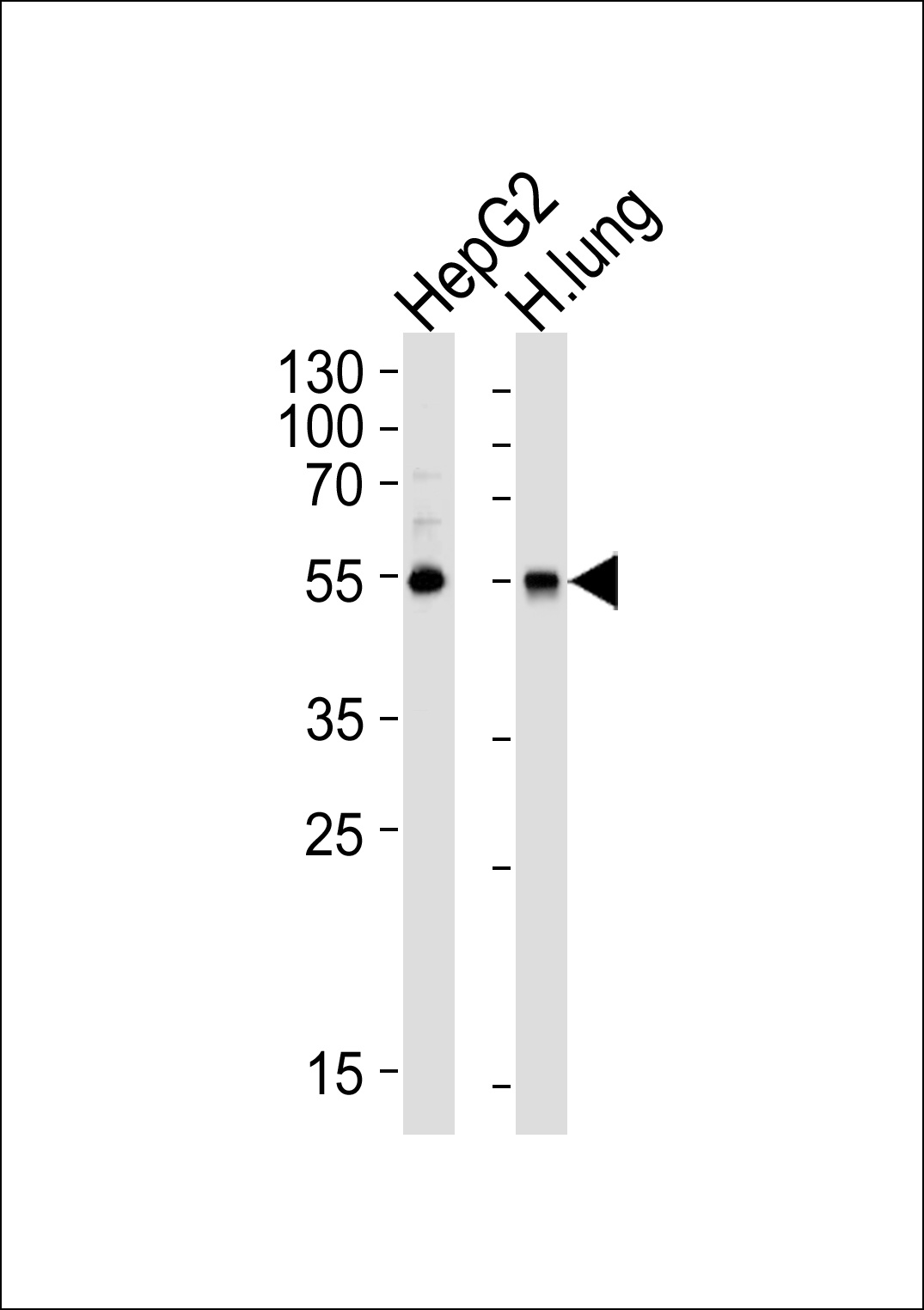
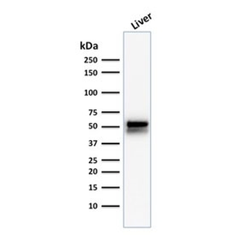
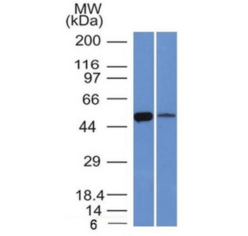
Western blot analysis of lysates from HepG2 cell line and human lung tissue lysate (from left to right), using ALDH1A1 Antibody at 1:1000 at each lane.
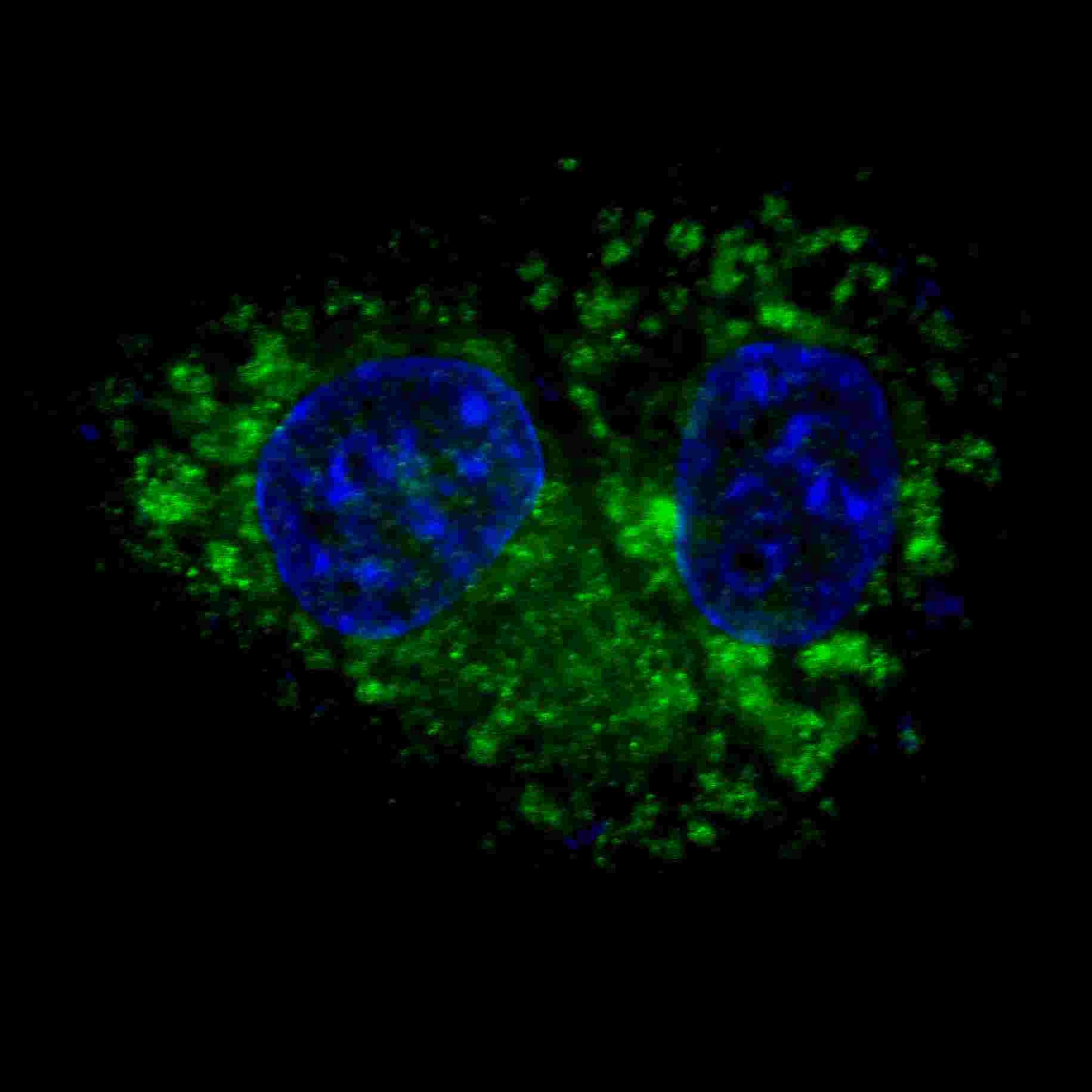
Fluorescent confocal image of HepG2 cells stained with ALDH1A1 antibody. HepG2 cells were fixed with 4% PFA (20 min), permeabilized with Triton X-100 (0.2%, 30 min). Cells were then incubated with ALDH1A1 primary antibody (1:100, 2 h at room temperature). For secondary antibody, Alexa Fluor 488 conjugated donkey anti-rabbit antibody (green) was used (1:1000, 1h). Nuclei were counterstained with Hoechst 33342 (blue) (10 ug/ml, 5 min). ALDH1A1 immunoreactivity is localized to the cytoplasm of HepG2 cells.
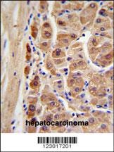
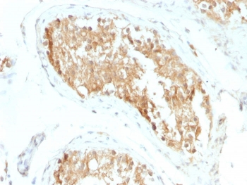
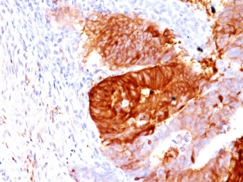
ALDH1A1 Antibody IHC analysis in formalin fixed and paraffin embedded human hepatocarcinoma followed by peroxidase conjugation of the secondary antibody and DAB staining.
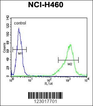
Flow cytometric analysis of NCI-H460 cells (right histogram) compared to a negative control cell (left histogram). FITC-conjugated goat-anti-rabbit secondary antibodies were used for the analysis.
- Item 1 of 10
Retinal dehydrogenase 1 ALDH1A1 Antibody [orb570330]
ELISA, FC, ICC, IF, IHC, WB
Human, Mouse, Rat
Rabbit
Polyclonal
Unconjugated
10 μg, 100 μg - Item 1 of 6
ALDH1A1 antibody [orb48279]
ICC, IF, IHC, WB
Human, Mouse, Rat
Polyclonal
Unconjugated
200 μl, 100 μl, 50 μl - Item 1 of 5
ALDH1A1 Antibody [orb1268148]
FC, IF, IHC-P, WB
Monkey, Mouse
Human
Rabbit
Polyclonal
Unconjugated
400 μl - Item 1 of 5
- Item 1 of 5
ALDH1A1 Antibody / Acetaldehyde dehydrogenase 1 [orb639650]
IHC-P, WB
Human
Mouse
Monoclonal
Unconjugated
100 μg, 20 μg


Submit a review
Filter by Rating

- 5 stars

- 4 stars
- 3 stars
- 2 stars
- 1 stars
