You have no items in your shopping cart.
Cart summary
Item 1 of 6
Item 1 of 6
AKT3 FITC Antibody
Catalog Number: orb344535

| Catalog Number | orb344535 |
|---|---|
| Category | Antibodies |
| Description | AKT3 FITC antibody (FITC) |
| Species/Host | Mouse |
| Clonality | Monoclonal |
| Clone Number | 25F6.F6.D8 |
| Tested applications | DOT, ELISA |
| Reactivity | Human, Mouse, Rat |
| Isotype | IgG1 |
| Immunogen | Anti-AKT3 Antibody was prepared from tissue culture supernatant by Protein A affinity chromatography using a synthetic peptide corresponding to internal residues of human AKT3 protein. |
| Antibody Type | Primary Antibody |
| Concentration | 1.0 mg/mL |
| Dilution range | ELISA: User Optimized, FC: User Optimized, IHC: User Optimized, IF: User Optimized, WB: User Optimized |
| Form/Appearance | Lyophilized |
| Purity | Anti-AKT3 antibody is directed against human AKT3. The antibody detects both unphosphorylated and phosphorylated forms of the protein. Anti-AKT3 antibody was purified from tissue culture by Protein A chromatography. Cross reactivity with AKT3 from other species has not been determined, however, the sequence of the immunogen shows 100% identity to human, mouse, and rat, therefore, cross reactivity is expected. Cross-reactivity with AKT2 and AKT has not been determined. |
| Conjugation | FITC |
| UniProt ID | Q9Y243 |
| NCBI | NP_001193658.1 |
| Storage | Store vial at 4° C prior to restoration. Restore with deionized water (or equivalent). For extended storage aliquot contents and freeze at -20° C or below. Avoid cycles of freezing and thawing. Centrifuge product if not completely clear after standing at room temperature. This product is stable for several weeks at 4° C as an undiluted liquid. Dilute only prior to immediate use. Expiration date is one (1) year from date of restoration. |
| Buffer/Preservatives | 0.01% (w/v) Sodium Azide |
| Alternative names | Mouse anti-AKT3 antibody FITC conjugation, fluores Read more... |
| Note | For research use only |
| Application notes | Anti-AKT3 FITC Antibody has been tested by ELISA and dot blot and is suitable for Flow Cytometry, immunohistochemistry, and western blotting. Expect a band approximately 56 kDa in size corresponding to AKT3 protein by western blotting in the appropriate cell lysate or extract. This monoclonal antibody reacts with human AKT. Specific conditions for reactivity should be optimized by the end user. For immunohistochemistry we recommend the use of fresh frozen tissues. Attempts at staining paraffin-embedded formalin fixed tissues were negative. No pre-treatment of sample is required. |
| Expiration Date | 12 months from date of receipt. |
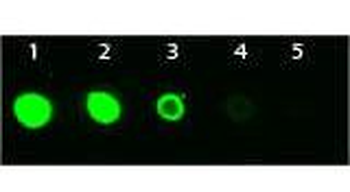
Dot Blot of Mouse anti-AKT3 Monoclonal Antibody Fluorescein Conjugated. Antigen: His-tagged AKT3. Load: Lane 1 - 100 ng Lane 2 - 33.3 ng Lane 3 - 11.1 ng Lane 4 - 3.70 ng Lane 5 - 1.23 ng. Primary antibody: n/a. Secondary antibody: Mouse anti-AKT3 Monoclonal Antibody Fluorescein Conjugated at 1:1000 for 60 min at RT. Block: orb348637 for 1 HR at RT.
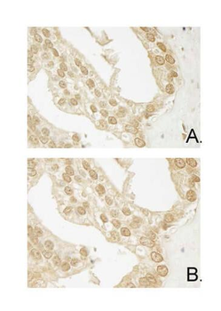
Immunohistochemistry of Mouse Anti-AKT3 antibody. Tissue: human prostate carcinoma. A) AKT-3 antibody produced using CELLine, B) AKT-3 antibody produced using roller bottle. Fixation: formalin fixed paraffin embedded. Antigen retrieval: not required. Primary antibody: AKT-3 antibody at 10 µg/ml for 1 h at RT. Secondary antibody: Peroxidase mouse secondary antibody at 1:10000 for 1 h at RT. Localization: AKT3 is nuclear and occasionally cytoplasmic. Staining: AKT3 as precipitated brown signal with hematoxylin purple nuclear counterstain.
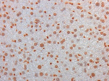
Immunohistochemistry of Mouse Monoclonal anti AKT3 Antibody in Mouse Embryonic Kidney. Tissue: Mouse Liver. Fixation: FFPE buffered formalin 10% conc. Ag Retrieval: Heat, Citrate pH6.2. Pressure Cooker. Primary antibody: anti-AKT3 at 2 ug/ml for 1.5 hour @ room Temp. Secondary Ab: MOUSE ON MOUSE HRP POLYMER 45" RT.
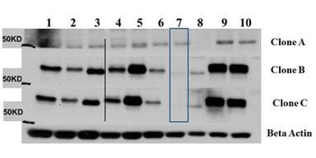
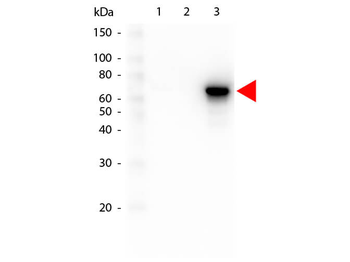
Western Blot of Mouse Anti-AKT3 antibody. Lane 1: C2C12. Lane 2: MEF#1. Lane 3: MEF#2. Lane 4: A549. Lane 5: Calu-1. Lane 6: PC3. Lane 7: HepG2. Lane 8: Jurkat. Lane 9: SKOV3. Lane 10: 293T. Load: 35 µg per lane. Primary antibody: AKT-3 antibody at 1:1000 for overnight at 4°C. Secondary antibody: Anti mouse secondary antibody at 1:20000 for 1 h at RT. Block: 5% BLOTTO overnight at 4°C. Predicted/Observed size: 56 kDa for AKT3.
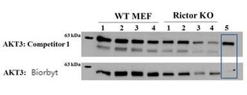
Western Blot of Mouse anti-AKT3 antibody. Lane 1: Control. Lane 2: Rapa. Lane 3: T50. Lane 4: T250. Lane 5: Control. Lane 6: Rapa. Lane 7: T50. Lane 8: T250. Lane 9: AKT3 null. Load: 35 µg per lane. Primary antibody: AKT-3 antibody at 1:1000 for overnight at 4°C. Secondary antibody: Anti mouse secondary antibody at 1:20000 for 1 h at RT. Block: 5% BLOTTO overnight at 4°C. Predicted/Observed size: 56 kDa for AKT3.
Western Blot of Mouse anti-AKT3 antibody. Lane 1: GST Tagged recombinant AKT1. Lane 2: GST Tagged recombinant AKT2. Lane 3: GST Tagged recombinant AKT3. Load: 25 ng per lane. Primary antibody: AKT3 antibody at 1:1000 for overnight at 4°C. Secondary antibody: Peroxidase mouse secondary antibody at 1:40000 for 30 min at RT. Block: orb348637 for 30 min at RT. Predicted/Observed size: 78 kDa for AKT3. Other band(s): none.

AKT3 Rabbit Polyclonal Antibody (FITC) [orb9635]
IF
Canine, Equine, Gallus, Rabbit, Sheep
Human, Mouse, Rat
Rabbit
Polyclonal
FITC
100 μlPhospho-AKT3 (Ser472) Rabbit Polyclonal Antibody (FITC) [orb9638]
IF
Bovine, Canine, Gallus, Porcine, Rabbit, Sheep
Human, Mouse, Rat
Rabbit
Polyclonal
FITC
100 μlPhospho-AKT1 + AKT2 + AKT3 (Thr308+Thr309+Thr305) Rabbit Polyclonal Antibody (FITC) [orb9628]
FC, ICC, IF
Bovine, Canine, Gallus, Porcine, Rabbit, Sheep
Human, Mouse, Rat
Rabbit
Polyclonal
FITC
100 μl
