You have no items in your shopping cart.
Cart summary
Item 1 of 5
Item 1 of 5
AKT1 FITC Antibody
Catalog Number: orb344537

| Catalog Number | orb344537 |
|---|---|
| Category | Antibodies |
| Description | AKT1 FITC antibody (FITC) |
| Species/Host | Mouse |
| Clonality | Monoclonal |
| Clone Number | 5E5.F5.D7 |
| Tested applications | ELISA, WB |
| Reactivity | Human, Mouse, Rat |
| Isotype | IgG2a |
| Immunogen | Anti-AKT1 Antibody was produced in mice by repeated immunizations with a synthetic peptide corresponding to internal residues of human AKT1 protein followed by monoclonal development. |
| Antibody Type | Primary Antibody |
| Concentration | 1.0 mg/mL |
| Dilution range | ELISA: User Optimized, FC: User Optimized, IHC: User Optimized, IF: User Optimized, WB: User Optimized |
| Form/Appearance | Lyophilized |
| Purity | Anti-AKT1 antibody is directed against human AKT1. The antibody detects both unphosphorylated and phosphorylated forms of the protein. Anti-AKT1 antibody was purified from ascites by Protein A chromatography. Cross reactivity with AKT1 from other species has not been determined, however, the sequence of the immunogen shows 85% identity to mouse and 92% identity with rat, therefore, cross reactivity is expected. |
| Conjugation | FITC |
| UniProt ID | P31749 |
| NCBI | NP_001014431.1 |
| Storage | Store vial at 4° C prior to restoration. Restore with deionized water (or equivalent). For extended storage aliquot contents and freeze at -20° C or below. Avoid cycles of freezing and thawing. Centrifuge product if not completely clear after standing at room temperature. This product is stable for several weeks at 4° C as an undiluted liquid. Dilute only prior to immediate use. Expiration date is one (1) year from date of restoration. |
| Buffer/Preservatives | 0.01% (w/v) Sodium Azide |
| Alternative names | mouse anti-AKT1 antibody FITC conjugation, fluores Read more... |
| Note | For research use only |
| Application notes | Anti-AKT1 FITC Antibody has been tested by ELISA and western blot and is suitable for Flow Cytometry, immunohistochemistry, and western blotting. Expect a band approximately 56 kDa in size corresponding to AKT1 protein by western blotting in the appropriate cell lysate or extract. This monoclonal antibody reacts with human AKT. Specific conditions for reactivity should be optimized by the end user. For immunohistochemistry we recommend the use of fresh frozen tissues. Attempts at staining paraffin-embedded formalin fixed tissues were negative. No pre-treatment of sample is required. |
| Expiration Date | 12 months from date of receipt. |

ELISA of Mouse Monoclonal anti-AKT1 antibody. Antigen: GST AKT1, GST AKT2, GST AKT3. Coating amount: starting from 50 ng/well. Primary antibody: Mouse monoclonal anti-AKT1 antibody at 100 ng/well. Dilution series: 2-fold. Mid-point concentration: 3 ng/mL Mouse monoclonal anti-AKT1 antibody. Secondary antibody: Peroxidase mouse secondary antibody at 1:10000. Substrate: TMB.

Flow Cytometry of Mouse anti-AKT1 antibody. Cells: LNCap Cells. Stimulation: none. Primary antibody: Allophycocyanin AKT1 antibody at 1.0 µg/ml for 20 min at 4°C.
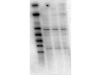
Western Blot of Mouse anti-AKT1 antibody. Lane 1: GST-AKT1. Lane 2: GST-AKT2. Lane 3: GST-AKT3. Lane 4: Molecular Weight Marker. Load: 25 ng per lane. Primary antibody: AKT1 antibody at 1:1000 for overnight at 4°C. Secondary antibody: Mouse secondary antibody at 1:40000 for 30 min at RT. Block: 5% BLOTTO overnight at 4°C. Predicted/Observed size: 78 kDa for AKT1. Other band(s): none.
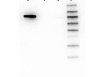
Western Blot of Mouse Anti-AKT1 antibody. Lane 1: Molecular Weight Marker. Lane 2: LnCap lysate (p/n orb348694). Lane 3: Jurkat lysate (p/n orb348674). Lane 4: MDA-MB 468 lysate (p/n orb348690). Load: 5 µg per lane. Primary antibody: AKT1 antibody at 1:1000 for overnight at 4°C. Secondary antibody: Mouse secondary antibody at 1:20000 for 45 min at RT. Block: 5% BLOTTO overnight at 4°C. Predicted/Observed size: 56 kDa for AKT1.
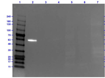
Western Blot of Mouse Anti-AKT1 Fluorescein Conjugated Antibody. Lane 1: Opal Prestained Molecular Weight Marker. Lane 2: AKT1 Recombinant Protein (0.05 µg). Lane 3: AKT2 Recombinant Protein (0.05 µg). Lane 4: AKT3 Recombinant Protein (0.05 µg). Lane 5: HEPG2 Whole Cell Lysate (20 µg). Lane 6: C2C12 Whole Cell Lysate (20 µg). Lane 7: A549 Whole Cell Lysate (20 µg). Primary Antibody: Mouse Anti-AKT1 FITC at 1.0 µg/ml overnight at 2-8°C. Secondary Antibody: Rabbit Anti-Mouse HRP (p/n orb347506) at 1:40000 for 30 mins at RT. Blocking: Fluorescent Buffer (p/n orb348644) for 1hr at RT. Predicted MW: ~56kDa. Exposure: 0.5 sec.

AKT1S1 Rabbit Polyclonal Antibody (FITC) [orb190207]
IF
Bovine, Equine, Mouse, Porcine, Rat, Sheep
Human
Rabbit
Polyclonal
FITC
100 μlPhospho-AKT1S1 (Thr246) Rabbit Polyclonal Antibody (FITC) [orb9641]
IF
Bovine, Canine, Equine, Human, Mouse
Rat
Rabbit
Polyclonal
FITC
100 μlAKT1 Rabbit Polyclonal Antibody (FITC) [orb2081280]
WB
Bovine, Canine, Goat, Human, Mouse, Porcine, Rat, Sheep
Rabbit
Polyclonal
FITC
100 μl
