You have no items in your shopping cart.
Cart summary
Item 1 of 7
Item 1 of 7
AKT1 antibody
Catalog Number: orb750474

| Catalog Number | orb750474 |
|---|---|
| Category | Antibodies |
| Description | AKT1 antibody |
| Species/Host | Rabbit |
| Clonality | Polyclonal |
| Tested applications | ELISA, FC, IF, IHC, WB |
| Reactivity | Gallus, Human, Mouse, Rat |
| Isotype | Antiserum |
| Immunogen | AKT Antibody was produced from whole rabbit serum prepared by repeated immunizations with a synthetic peptide R-P-H-F-P-Q-F-S-Y-S-A-S-G-T-A corresponding to the C-terminus (460-480) of human AKT proteins conjugated to KLH using maleimide. A residue of cysteine was added to the amino terminal end to facilitate coupling. A BLAST analysis was used to suggest reactivity with this protein from rat, mouse, and chicken based on 100% homology for the immunogen sequence. |
| Concentration | 85 mg/mL |
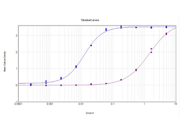
| Dilution range | ELISA: 1:2,000 - 1:10,000, FC: User Optimized, IHC: 1:500 - 1:2,000, IF: 1:100 - 1:1,000, WB: 1:500 - 1:2,000 |
| Form/Appearance | Liquid (sterile filtered) |
| Purity | This product was prepared from monospecific antiserum by a delipidation and defibrination. Pan Anti-AKT Antibody reacts with the AKT from human tissues. Based on sequence we expect this antibody to react as well with rat, mouse, and chicken AKT. |
| Conjugation | Unconjugated |
| UniProt ID | P31749 |
| NCBI | 62241011 |
| Storage | Store Anti-Akt antibody at -20° C prior to opening. Aliquot contents and freeze at -20° C or below for extended storage. Avoid cycles of freezing and thawing. Centrifuge product if not completely clear after standing at room temperature. This product is stable for several weeks at 4° C as an undiluted liquid. Dilute only prior to immediate use. |
| Buffer/Preservatives | 0.01% (w/v) Sodium Azide |
| Alternative names | rabbit anti-AKT antibody, RAC-PK-alpha, Protein ki Read more... |
| Note | For research use only |
| Application notes | Anti-AKT Antibody has been tested in Western Blot, Immunohistochemistry (Formalin-fixed paraffin-embedded sections), and Immunofluorescence (paraformaldehyde-fixed primary cardiomyocyte cultures). Expect a band at ~55.7kDa in 3T3 whole cell lysate or other appropriate cell lysates or tissues in western blot. Although not tested, this antibody would be useful in flow cytometry. Researchers should determine optimal titers for applications that are not stated below. |
| Expiration Date | 12 months from date of receipt. |
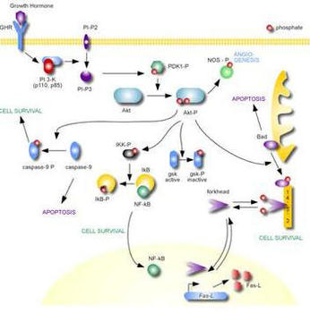
AKT Metabolic Pathway.
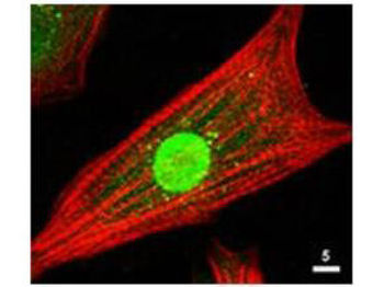
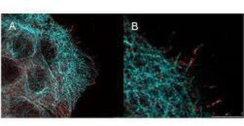
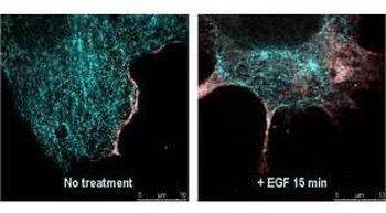
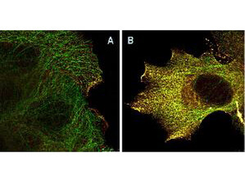
Immunofluorescence Microscopy of Rabbit Anti-AKT Antibody. Tissue: neonatal rat cardiomyocytes. Fixation: 0.5% PFA. Antigen retrieval: not required. Primary antibody: AKT antibody at 1:80 dilution for 1 h at RT. Secondary antibody: Texas-red™ conjugated rabbit secondary antibody at 1:10000 for 45 min at RT. Localization: AKT is nuclear. Staining: Anti-AKT staining appears green. Actin filaments are labeled red using a Texas-red™ conjugated phalloidin.
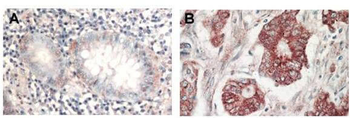
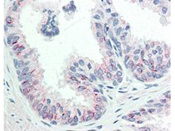
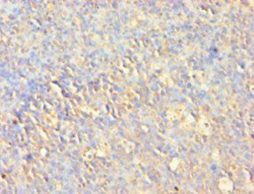
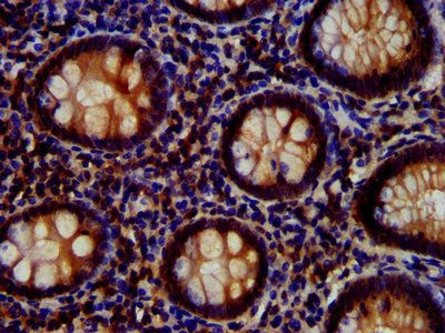
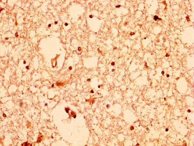
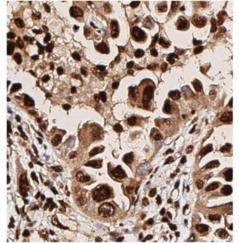
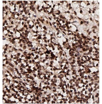
Immunohistochemistry of Rabbit Anti-AKT antibody. Tissue: (A) normal colon tissue, (B) colon tumor tissue. Fixation: formalin fixed paraffin embedded. Antigen retrieval: not required. Primary antibody: AKT antibody at 1:1000 dilution for 1 h at RT. Secondary antibody: Peroxidase rabbit secondary antibody at 1:10000 for 45 min at RT. Localization: AKT is nuclear. Staining: AKT as precipitated red signal with hematoxylin purple nuclear counterstain.
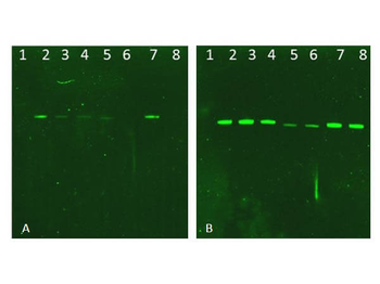
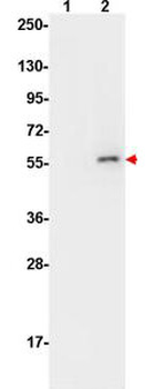
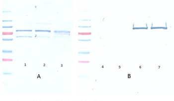
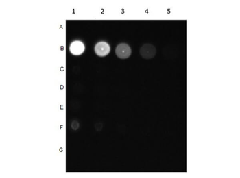
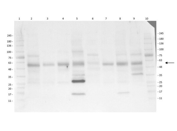
Western Blot of Rabbit AKT Antibodies. Lane 1: NIR MW protein ladder. Lane 2: AKT1, recombinant: orb346473. Lane 3: AKT1, phosphatase-treated: orb346472. Lane 4: AKT1, mutant T308A/S473A: orb346474. Lane 5: AKT2, recombinant: orb346475. Lane 6: AKT2, phosphatase-treated: orb346470. Lane 7: AKT3, recombinant: orb346476. Lane 8: AKT3, phosphatase-treated: orb346471. Load: 50 ng per lane. Blot A: orb345379 Anti-Akt pT308 used at 1:2270, Blot B: orb750474 Anti-Akt used 1:1000.
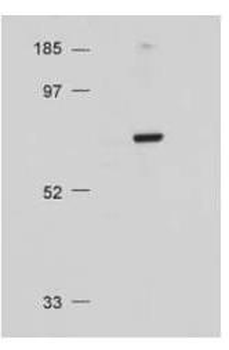
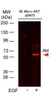
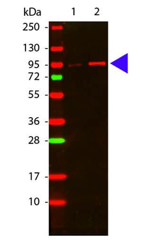
Western Blot of Rabbit Anti-AKT antibody. Lane 1: Molecular Weight. Lane 2: NIH/3T3 whole cell lysate. Load: 20 µg lysate per lane. Primary antibody: Anti-AKT antibody at 1:500 for overnight at 4°C. Secondary antibody: HRP conjugated GT-a-Rabbit IgG (orb347654) at 1:10000 preceded color development. Block: MOPS buffer overnight at 4°C. Predicted/Observed size: 56 kDa, 56 kDa for AKT. Other band(s): none.
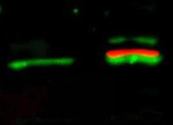
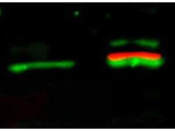
Western Blot of simultaneous detection of unphosphorylated and phosphorylated Rabbit Anti-AKT antibody. Lane 1: unstimulated NIH/3T3 lysates contain inactive unphosphorylated Akt1, green band. Lane 2: PDGF stimulated NIH/3T3 lysate contains both inactive (green band) and activated phosphorylated Akt1 (red band). Load: 35 µg per lane. Primary antibody: rabbit anti-Akt (pan) and mouse anti-Akt pS473 specific antibodies at 1:1000 for overnight at 4°C. Secondary antibody: DyLight™ 549 conjugated anti-rabbit IgG (green) and DyLight™ 649 conjugated anti-mouse IgG (red) secondary antibodies at 1:10000 for 45 min at RT. Block: 5% BLOTTO overnight at 4°C.
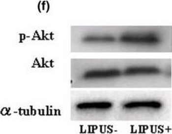
Western blotting analysis. (a) Type-II collagen. (b) Type-IX collagen. (c) Focal adhesion kinase (FAK) and phosphorylated FAK (p-FAK). (d) Paxillin and phosphorylated Paxillin (p-Paxillin). (e) Mitogen-activated protein kinase (MAPK) and phosphorylated MAPK (p-MAPK). There are no evident differences in the expression levels of total MAPK and p-MAPK between the two groups. (f) Akt and phosphorylated Akt (p-Akt). There were no differences found in the intensity the total Akt expression between the two groups, but p-Akt was found at higher levels in the LIPUS group (US+) in comparison with the control group (US-). (g) Cyclin B1 and cyclin D1. (h) Changes of proliferating cell nuclear antigen (PCNA) using MEK1 inhibitor (PD98059) and phosphatidylinositol 3-OH kinase (PI3K) inhibitor (LY294002). Chondrocytes were pretreated with MEK1 inhibitor (PD98059, 250 μM/ml) and PI3K inhibitor (LY294002, 250 μM/ml) for 12 hours and 24 hours followed by stimulation with LIPUS for 20 minutes. Each sample was harvested 2 hours after LIPUS stimulation and the influence of these inhibitors was judged in western blotting analysis of the expression of PCNA.
- Item 1 of 9
Akt (phospho-S473) antibody [orb344403]
ELISA, FC, IF, IHC, IP, Multiplex Assay, WB
Human, Monkey, Mouse, Rat
Mouse
Monoclonal
Unconjugated
100 μg - Item 1 of 9
Akt (phospho-S473) antibody [orb344404]
ELISA, FC, IF, IHC, IP, Multiplex Assay, WB
Human, Monkey, Mouse, Rat
Mouse
Monoclonal
Unconjugated
25 μl - Item 1 of 8
- Item 1 of 8
Akt (phospho-T308) antibody [orb345379]
DOT, ELISA, FC, IHC, WB
Human, Mouse, Rat
Rabbit
Polyclonal
Unconjugated
100 μg - Item 1 of 8
AKT (phospho-T308) antibody [orb420308]
DOT, ELISA, FC, IHC, WB
Human, Mouse, Rat
Rabbit
Polyclonal
Unconjugated
25 μl


Submit a review
Filter by Rating

- 5 stars

- 4 stars
- 3 stars
- 2 stars
- 1 stars
