You have no items in your shopping cart.
Cart summary
Item 1 of 6
Item 1 of 6
AKT1 antibody
Catalog Number: orb344524

| Catalog Number | orb344524 |
|---|---|
| Category | Antibodies |
| Description | AKT1 antibody |
| Species/Host | Mouse |
| Clonality | Monoclonal |
| Clone Number | 14E5.A2.B2.H9 |
| Tested applications | ELISA, FC, IHC, WB |
| Reactivity | Human, Mouse |
| Isotype | IgG2a |
| Immunogen | Anti-AKT1 Antibody was produced by repeated immunizations with a synthetic peptide corresponding to internal residues of human AKT1 protein. |
| Concentration | 1.0 mg/ml |
| Dilution range | ELISA: 1:2,000 - 1:10,000, FC: User Optimized, IHC: 20 µg/mL, WB: 1:1000 |
| Form/Appearance | Liquid (sterile filtered) |
| Purity | Anti-AKT1 antibody is directed against human AKT1. The antibody detects both unphosphorylated and phosphorylated forms of the protein. Anti-AKT1 antibody was purified from ascites by Protein A chromatography. Cross reactivity with AKT1 from other species has not been determined, however, the sequence of the immunogen shows 85% identity to mouse and 92% identity with rat, therefore, cross reactivity is expected. |
| Conjugation | Unconjugated |
| UniProt ID | P31749 |
| Storage | Store vial at -20° C prior to opening. Aliquot contents and freeze at -20° C or below for extended storage. Avoid cycles of freezing and thawing. Centrifuge product if not completely clear after standing at room temperature. This product is stable for several weeks at 4° C as an undiluted liquid. Dilute only prior to immediate use. |
| Buffer/Preservatives | None |
| Alternative names | mouse anti-AKT1 antibody, AKT-1, PKB antibody, PKB Read more... |
| Note | For research use only |
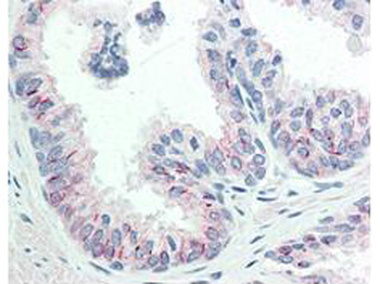
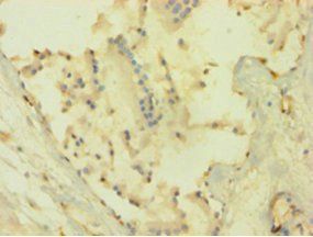
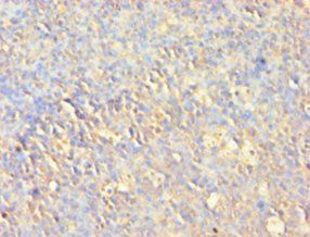
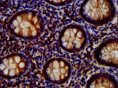
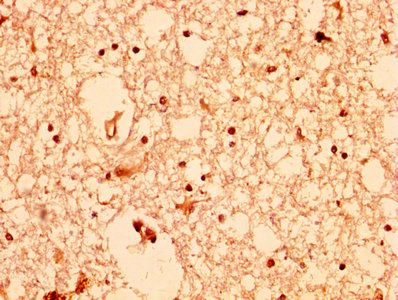
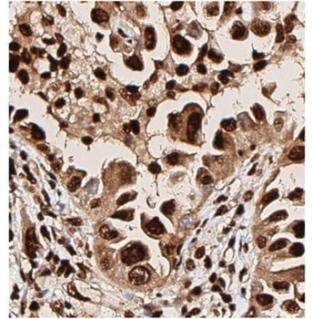
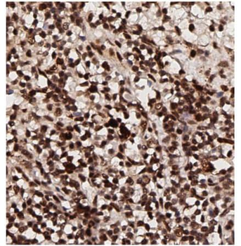
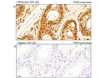
| Application notes | Anti-AKT1 Antibody has been tested in ELISA, SDS-Page, flow cytometry, and western blotting. This antibody is suitable for immunoprecipitation and immunohistochemistry. Expect a band approximately 56 kDa in size corresponding to AKT1 protein by western blotting in the appropriate cell lysate or extract. This monoclonal antibody reacts with human AKT. Specific conditions for reactivity should be optimized by the end user. For immunohistochemistry we recommend the use of fresh frozen tissues. Attempts at staining paraffin-embedded formalin fixed tissues were negative. No pre-treatment of sample is required. |
| Expiration Date | 12 months from date of receipt. |
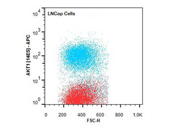
Flow Cytometry of Mouse anti-AKT1 antibody. Cells: LNCap Cells. Stimulation: none. Primary antibody: Allophycocyanin AKT1 antibody at 1.0 µg/mL for 20 min at 4°C.
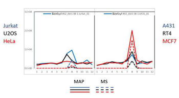
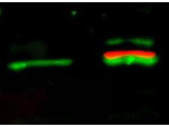
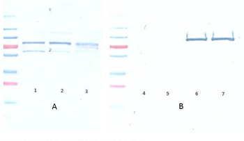
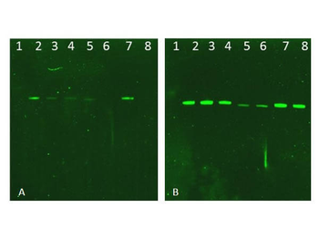
PAGE-MAP (microsphere affinity proteomics) of Mouse Anti-AKT1 Antibody. (Catalog Number: orb344523). Antibody array western blot binding of gelfree size separated fractions of multiple lysates (solid lines) and shotgun mass spectroscopy identification (dashed lines) of the target band run in parallel correlate confirming the specificity of this antibody against AKT1.
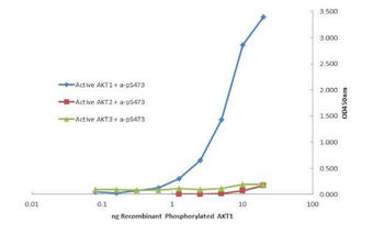
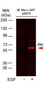
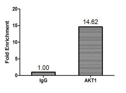
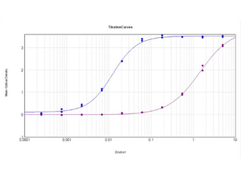
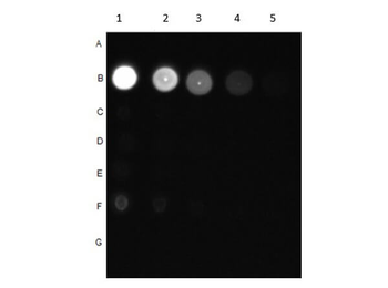
Plate was coated with monoclonal anti AKT1 antibody (capture antibody) followed by incubation with recombinant AKT1 (p/n orb346473), AKT2 (p/n orb346474), AKT3 (p/n orb346475) proteins. Binding was detected with biotinylated monoclonal anti-AKT pS473. The signal shows specificity of the monoclonal anti-AKT1 antibody to recombinant isoform AKT1 protein and not the isoform 2 and 3.
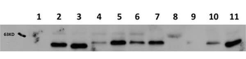
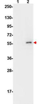
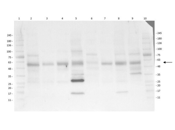
Western Blot of Mouse Anti-AKT1 antibody. Lane 1: AKT-1 Null. Lane 2: WT. Lane 3: MEF #1. Lane 4 : A549. Lane 5: Calu-1. Lane 6: PC-3 (p/n orb692718). Lane 7: HepG2 (p/n orb348735). Lane 8: Jurkat (p/n orb348674). Lane 9: SKOV3. Lane 10: HEK293T. Lane 11: C2C12 (p/n orb348725). Load: 20 ug per lane. Primary antibody: AKT1 antibody at 1:1000 for overnight at 4°C. Secondary antibody: Peroxidase mouse secondary antibody at 1:40000 for 30 min at RT. Block: orb348637 for 30 min at RT. Predicted/Observed size: 56 kDa for AKT1. Other band(s): none.
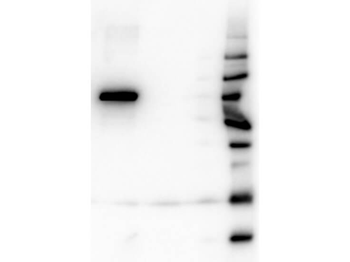
Western Blot of Mouse Anti-AKT1 antibody. Lane 1: GST Tagged recombinant AKT1. Lane 2: GST Tagged recombinant AKT2. Lane 3: GST Tagged recombinant AKT3. Load: 25 ng per lane. Primary antibody: AKT1 antibody at 1:1000 for overnight at 4°C. Secondary antibody: Peroxidase mouse secondary antibody at 1:40000 for 30 min at RT. Block: orb348637 for 30 min at RT. Predicted/Observed size: 78 kDa for AKT1. Other band(s): none.
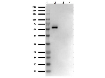
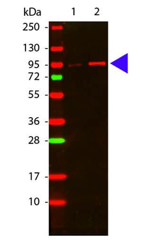
Western Blot of Mouse Anti-AKT1 Antibody. Lane 1: Opal Prestained Molecular Weight Protein. Lane 2: AKT1 protein (p/n orb346473). Lane 3: AKT2 protein (p/n orb346474). Lane 4: AKT3 protein (p/n orb346475). Load: 50 ng. Blocking: BlockOut Buffer (p/n orb348644) for 30 min at RT. Primary Antibody: Anti-AKT1 at 1 ug/ml o/n at 4°C. Secondary Antibody: Rabbit Anti-Mouse IgG HRP (p/n orb347506) at 1:40000 in orb348644 for 30 min at RT.
- Item 1 of 9
Akt (phospho-S473) antibody [orb344403]
ELISA, FC, IF, IHC, IP, Multiplex Assay, WB
Human, Monkey, Mouse, Rat
Mouse
Monoclonal
Unconjugated
100 μg - Item 1 of 9
Akt (phospho-S473) antibody [orb344404]
ELISA, FC, IF, IHC, IP, Multiplex Assay, WB
Human, Monkey, Mouse, Rat
Mouse
Monoclonal
Unconjugated
25 μl - Item 1 of 8
- Item 1 of 8
Akt (phospho-T308) antibody [orb345379]
DOT, ELISA, FC, IHC, WB
Human, Mouse, Rat
Rabbit
Polyclonal
Unconjugated
100 μg - Item 1 of 8
AKT (phospho-T308) antibody [orb420308]
DOT, ELISA, FC, IHC, WB
Human, Mouse, Rat
Rabbit
Polyclonal
Unconjugated
25 μl


Submit a review
Filter by Rating

- 5 stars

- 4 stars
- 3 stars
- 2 stars
- 1 stars
